Search for more information
3D/4D imaging on the LX8 Abdomen, OB/GYN, Cardiac Screening, Vascular, Breast
2 years
Up/Down, Left/Right, Angular variation, Reverse

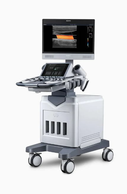

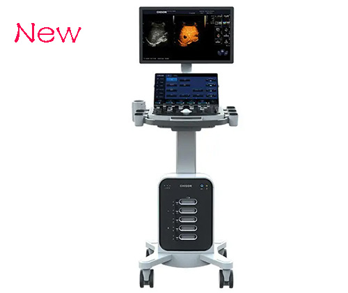
EDAN Acclarix LX8 Diagnostic Ultrasound System With Trolley Cart
Versatility & Innovation Meet Value & Performance
When it comes to power, performance, innovation and flexibility - there is one ultrasound system that delivers more. Designed specifically to address the challenges of busy ultrasound environments, the Acclarix LX8 Ultrasound System features a host of design breakthroughs that make day-to-day operation easy, fast and intuitive.

Features
3D/4D imaging on the LX8 system includes a complete suite of rendering and editing tools ensuring maximum results
Adaptive color Doppler provides exquisite sensitivity and detail resolution with minimal user optimization required as seen in this normal kidney
Tissue Adaptive Imaging with the L17-7HD transducer delivers exceptional contrast and detail resolution in this evaluation of a thyroid lesion
Superb performance of color and spectral Doppler is demonstrated in this view of the hepatic vein confluence
Low maintenance
Easy to use
Enhanced service life
Advanced technology used
Durable nature

Unparalleled image quality with one key optimization for all modes and all applications
User customizable 10” touch screen with innovative gesture control user interface
Large 21” tilt-and-swivel, HD wide-screen anti-glare LCD monitor
Image Quality Without Compromise - Flexibility, versatility and user-friendly features are just the beginning.
The Acclarix LX8 system also delivers the brilliant clarity and stunning image quality required in fast-paced environments - from hospitals to private practices to imaging centers.
Motorized height adjustment with 20-cm travel delivers a flexible ergonomic scanning position
Customizable touch screen user interface to personalize your workflow
Four active transducer ports so the transducer you need is always ready to use
High fidelity, high-channel count architecture results in superb detail resolution, particularly when scanning at depth
Tissue Adaptive Imaging (TAI) continuously and automatically optimizes imaging allowing more focus on the patient.
In B-mode, TAI fine tunes multiple imaging parameters to provide the best possible image quality.
In color and spectral Doppler, TAI automatically adjusts for flow state providing improved continuity, border detection and fill-in.

Features
3D/4D imaging on the LX8 system includes a complete suite of rendering and editing tools ensuring maximum results
Adaptive color Doppler provides exquisite sensitivity and detail resolution with minimal user optimization required as seen in this normal kidney
Tissue Adaptive Imaging with the L17-7HD transducer delivers exceptional contrast and detail resolution in this evaluation of a thyroid lesion
Superb performance of color and spectral Doppler is demonstrated in this view of the hepatic vein confluence
Low maintenance
Easy to use
Enhanced service life
Advanced technology used
Durable nature
TAI- Tissue Adaptive Imaging- Proprietary image technology from Edan
eSRI- Imaging Elimination Speckle
3D/4D capability to automate volume editing with eFace feature
Panoramic Imagery
Optimization of one contact picture
Imaging of TDI Tissue Doppler
Automated measurement instruments such as Auto IMT and Auto OBT
A detailed set of functions for DICOM
Technology for Needle Visualization