Search for more information
B, B/B, 4b, B/M, M, Pw, B/C, B/C/D, B/D, Duplex.
ECG access, access for foot switch
2 years
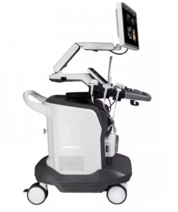
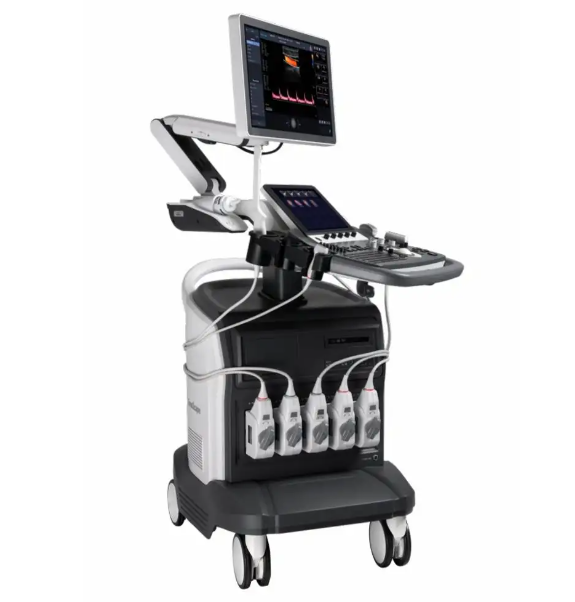
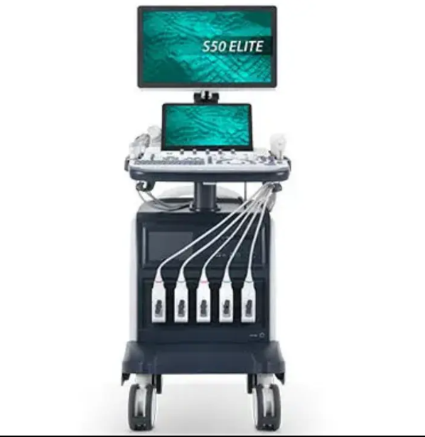
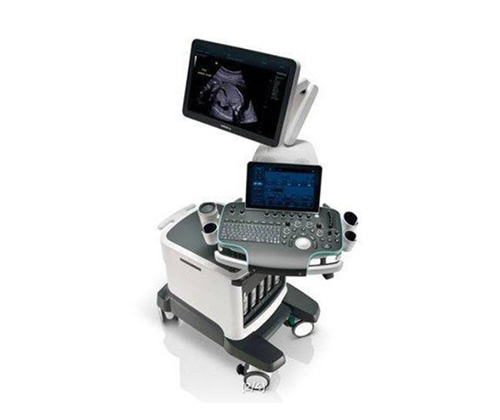
Sonoscape S50 Ultrasound Machine
Adhering to its great image quality, SonoScape’s newly released S50 carries a comprehensive, upgraded platform, to become an epoch-making ultrasound system. It is equipped with powerful single crystal transducers, remarkable 4D functions and intelligent workflow, which greatly helps to meet a wide variety of general imaging needs. Outfitted with extraordinary software and hardware, S50 is a good balanced design, internally and externally. The quick response touch screen, mode switching and storage operations deliver a new standard with clarity and flexibility.

C-xlasto Imaging
By understanding that tissue stiffness varies depending on the type of tissue, we can use this to easily find abnormalities and tumors within soft tissue. When applying consistent and repeated pressure with the transducer on soft tissue the system can quickly calculate the Strain Ratio of the region and give an Intelligent Strain Curve for a recording of applied pressure to assist with diagnosis of the abnormality. Predominately used only with linear probes, SonoScape’s new transvaginal probe for gynecological is breaking the mold and expanding elastography applications to non-linear probes.

Contrast Imaging
Contrast Imaging is the use of contrast agents that are small enough to go into the smallest of veins in the body and still provide a loud signal reflection for the ultrasound to pick up. Such a loud signal reflection allows the ultrasound to accurately display small veins in the body, blood profusion in different organs, and helps to measure blood flow. The S50 takes it a step farther by providing better resolution and deeper penetration, maximizing the short lifespan of the contrast agent bubbles by giving a clearer, more complete image of the desired organ.

Auto IMT
Auto IMT is used when determining the level of vascular sclerosis present in the patient by automatically tracing and calculating the thickness of the carotid vessels.
Tissue Doppler Imaging
Tissue Doppler Imaging allows you to quantitatively evaluate local myocardial movements and functions, with speed and strain/strain rate parameters.
Stress Echo
Stress echocardiography is the combination of 2D echocardiography with a physical, pharmacological or electrical stress of the patient. It also provides users with report management tools such as configurable template editor, multiple loops to select one for storage, wall motion scoring, stress echo report, etc

Wyposażenie:
21,5″ panoramiczny monitor LED FULL HD oraz wyjątkowo czuły i wygodny touchscreen o przekątnej 12”
5 złączy głowic obrazowych (w tym 4 aktywne i 1 postojowe)
1 złącze głowic nie obrazowych typu „ślepy doppler”
THI (tissueharmonicimaging) – tryb obrazowania harmonicznego
PHI (Pure Harmonic Imaging) – tryb obrazowania harmonicznego z inwersją fazy
Color Doppler i Power doppler
tryby obrazowania: B/2B/4B/M/CFM/PDI/DirPDI/PW/HPRF/CW
obrazowanie trapezoidalne
moduł kardiologiczny z pakietem pomiarowym i dopplerem CW (dostępny na głowicach typu PhasedArray)
anatomiczny M-mode
M-Tuning – optymalizacja obrazu jednym przyciskiem.