Search for more information
65L x 44W x 53H (cm) 38KG
Methodology: CCD Camera Resolution: 1280 x 1024 Live iris image: During alignment
$12300
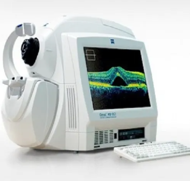
CARL ZEISS Cirrus HD-OCT 4000
Carl Zeiss Cirrus HD-OCT 4000 – The Cirrus HD-OCT is a non-contact, high resolution tomographic and biomicroscopic imaging device. It is indicated for in-vivo viewing, axial cross-sectional, and three-dimensional imaging and measurement of anterior and posterior ocular structures, including cornea, retina, retinal nerve fiber layer, ganglion cell plus inner plexiform layer, macula, and optic nerve head. The Cirrus normative databases are quantitative tools for the comparison of retinal nerve fiber layer thickness, macular thickness, ganglion cell plus inner plexiform layer thickness, and optic nerve head measurements to a database of normal subjects. The Cirrus HD-OCT is intended for use as a diagnostic device to aid in the detection and management of ocular diseases including, but not limited to, macular holes, cystoid macular edema, diabetic retinopathy, age-related macular degeneration, and glaucoma.
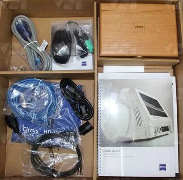
The ZEISS Certified Factory Direct Cirrus 4000 HD-OCT also includes the full Accessory Kit External Fixation Light
Test Eye
Mini Keyboard
Optical Mouse
Dust Cover
Metric 5A/250v Fuse
14 Ft. CAT 5e Network Cable
USB Cable
IEC 320 to NEMA 12 inch Power Cable
IEC 320 39in. Power Cable
Install Disc including Roxio Basic
Symbol Sheet
OCT Primer Guide
Quick Start Guide
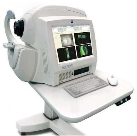
HD-OCT Imaging
Methodology: Spectral Domain OCT
Optical Source: superluminescent diode (SLD), 840 nm
Optical Power: < 725 μW at the cornea
Scan Speed: 27,000 A-scans per second
A-Scan depth: 2.0 mm (in tissue), 1024 points
Axial resolution: 5 μm (in tissue)
Transverse resolution: 15 μm (in tissue)
Zeiss Cirrus HD-OCT 4000 OCT Spectral Domain OCT HD Fundus Imaging
Methodology: Line scanning ophthalmoscope
Live Fundus Image: During alignment and during OCT scan
Optical Source: Superluminescent diode (SLD), 750 nm
Optical Power: < 1.5 mW at the cornea
Field of View: 36 degrees W x 30 degrees H
Frame rate: >20 Hz
Transverse resolution: 25 μm (in tissue)
Carl Zeiss Cirrus HD-OCT 4000 Iris Imaging
Methodology: CCD Camera
Resolution: 1280 x 1024
Live iris image: During alignment
Electrical, Physical and Environmental
Weight: 38kg (83 lbs)
Dimensions: 65L x 44W x 53H (cm)
Fixation: Internal, external
Internal fixation focus adjustment: -20D to +20D (diopters)
Input devices: Keyboard, mouse
Electrical rating (115V) Single Phase, 100/120V~ systems:50-60Hz, 5A
Fuse rating (115V) T 5A 250V
Electrical rating (230V) Single Phase, 220/240V~ systems:50-60 Hz, 2.5A
Fuse rating (230V) Fuse rating: T 5A 250V
Convenience Receptacle output ratings 115V~, 0.5 A Max, 50-60 Hz
Temperature (transport and storage) -40º to +70º C
Relative humidity (transport and storage) 10% to 100%, including condensation
Atmospheric pressure (transport and storage): 500 hPa to 1060 hPa
Temperature (operation) +10º to +35º C
Relative humidity (operation) 30% to 75%, excluding condensation
Atmospheric Pressure (operation) 700 hPa to 1060 hPa