Search for more information
True Color (with Red, Green and Blue channel separation) Autofluorescence-Green and Blue Infrared reflectance External eye image (ocular surface) Stereo
Fundus Camera Weight: 53 lbs Fundus Camera Dimensions (W x D x H): 15” × 22” × 27” inches Power Table Dimensions (W x D x H): 36” × 25” ×28” – 37” inches Power Table Weight: 84 lbs
6 Month warranty Power table Latest software Manual Dust cover
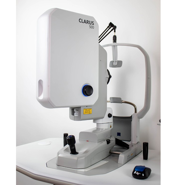
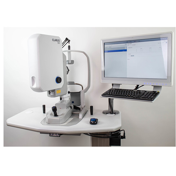
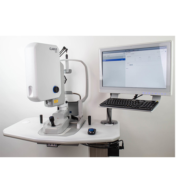
#ZEISS CLARUS 500 Imaging ultra-wide without compromise. The introduction of widefield retinal imaging has shown us that indications of disease are often located in the far periphery of the retina. CLARUS™ 500 from ZEISS, is an advanced fundus imaging system from ZEISS that provides True Color and high-resolution across an entire ultra-widefield image.

ZEISS CLARUS 500 captures clear Ultra-widefield images (200°-267°) and accurate images from the macula to the far periphery. Early indications of disease can often be subtle and difficult to see through direct observation or low resolution fundus imaging. Leveraging ZEISS optics, ZEISS CLARUS 500 is an ultra-widefield retinal camera that captures a high-resolution image down to 7 microns and generates images that closely resemble the coloration of the fundus as seen during clinical examination. The advent of widefield retinal imaging has shown us that indications of disease are often located in the far periphery of the retina. CLARUS 500 is the next-generation fundus imaging system from ZEISS that provides true color and high-resolution across an entire ultra-widefield image. The CLARUS 500 allows clinicians to track subtle changes in pathology over time. In addition to true color imaging, it also captures high resolution fundus autofluorescence (FAF) images–FAF-Blue and FAF-Green–and Infrared (IR) and external eye images. Along with an intuitive review software, the ultra-high-resolution of this next-generation retinal camera from ZEISS allows clinicians to manage change with confidence.

True Color:
Aids in diagnosis and documentation
CLARUS 500, the newest ultra-widefield retinal camera from ZEISS, allows clinicians to use color to their diagnostic advantage. It also generates images that closely resemble the coloration of the fundus as seen during clinical examination. Color fundus imaging can aid in the diagnosis and documentation of ocular disease, ensuing confidence when evaluating the optic disc, nevi and lesions where color is important.
Clarity:
From the macula to the periphery with ONE system
Legacy ultra-widefield imaging systems require doctors to maintain a traditional high-resolution fundus camera for optic nerve and macular disease diagnosis and management. CLARUS 500 from ZEISS is the first fundus imaging system to provide true color and clarity within an ultra-wide field of view, enabling clinicians to capture high-resolution fundus images from macula to the far periphery.

Comfort:
For a satisfying patient experience that ensures image integrity
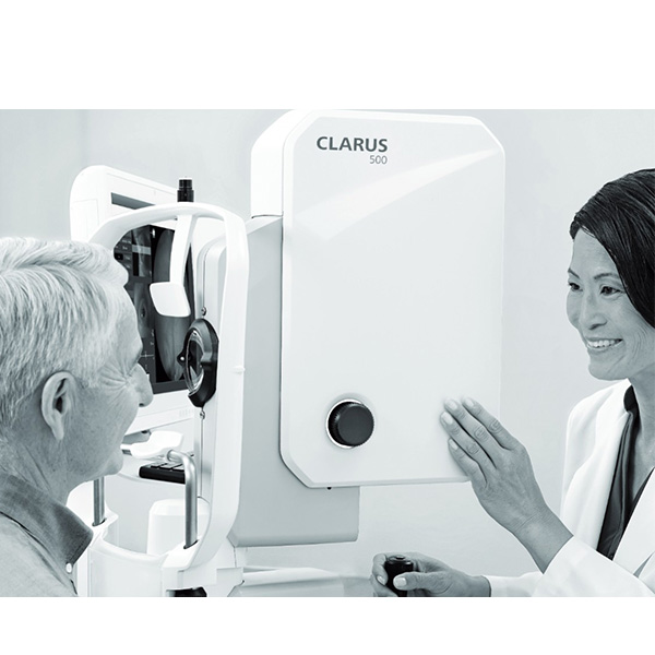
Simple, Stable and intuitive, ZEISS CLARUS 500 is an ultra-widefield retinal camera that has been purposefully designed to optimize each patient’s experience. By bringing the optics to the patient, ZEISS CLARUS 500 helps create a comfortable, satisfying patient experience that provides images free of obstructions, such as lids and lashes, and requires fewer recaptures. A stable and neutral head and chin rest brings the optics to the patient, facilitating an easier and more comfortable scan. The ability to swivel the device between the right and left eye helps technicians capture a high-quality image without realigning the patient.
Fewer recaptures with Live IR Preview
The Live IR Preview in ZEISS CLARUS 500 allows the technician to optimize alignment–intervene with lid and lash, and remove image artifacts before capturing an image. The result is fewer image captures for the patient and a more efficient imaging process for the practice.

Technical Specifications:
Parameters
Imaging Modes:
True Color (with Red, Green and Blue channel separation)
Autofluorescence-Green and Blue
Infrared reflectance
External eye image (ocular surface)
Stereo
Field of view (measured from the center of the eye):
Widefield (one image)- 133˚
Ultra-widefield (two images)- 267°
Montage (up to six images)- 133˚
Resolution: 7.3 µm
Minimum Pupil Diameter: 2.5 mm
Working Distance: 25 mm (patient’s eye to front lens)
Compensation for ametropia: – 24 D to + 20 D continuous
Light sources (LED): Red (585-640 nm), Green (500-585 nm), Blue (435-500 nm), Infrared laser diode (785 nm)
Automatic Operations:

Auto-focus
Auto-gain
Auto Montage: up to 267º Image Montage (up to six images)
Auto-laterality
Instrument Specifications:
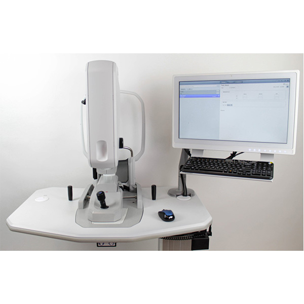
Fundus Camera Weight: 53 lbs
Fundus Camera Dimensions (W x D x H): 15” × 22” × 27” inches
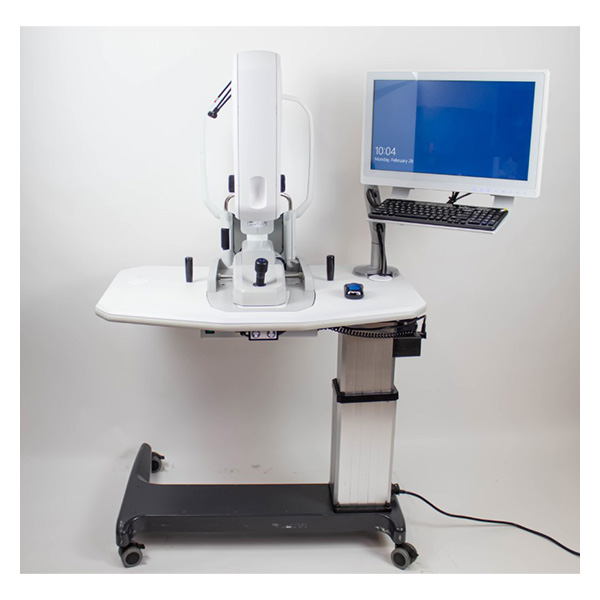
Power Table Dimensions (W x D x H): 36” × 25” ×28” – 37” inches
Power Table Weight: 84 lbs
This Zeiss Clarus 500 ultra-widefield fundus camera comes equipped with:
6 Month warranty
Power table
Latest software
Manual
Dust cover