
Search for more information
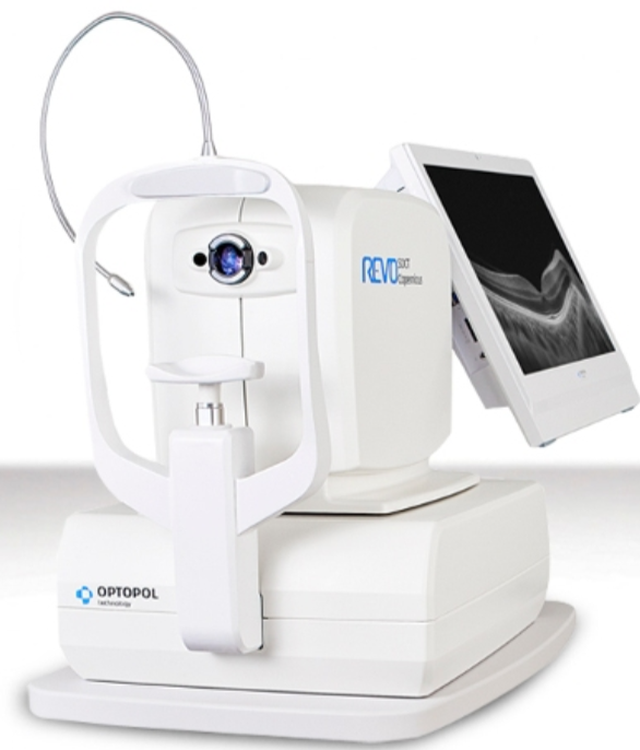
Fully automatic. Alignment, focus, and acquisition are fully automated. Extremely small dimensions and weights, like an auto refractometer. Scans in mioses up to 12mm. Retina and disc are analyzed thanks to a single scan. Front segment without any additional lens. It is possible to scan from both iridocorneal angles. Connection to any PC thanks to a single USB cable.
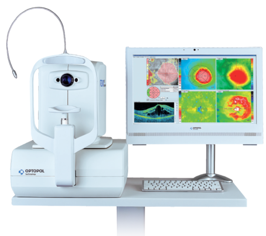
Analysis of 8 retinal layers with the regulatory database. Glaucoma analysis with nerve fiber thickness and Gangliari cell analysis with the regulatory database. Pachymetric and iridocorneal angle analysis. Methods of comparisons and follow-up of exams and pathologies. Optional angiography module.
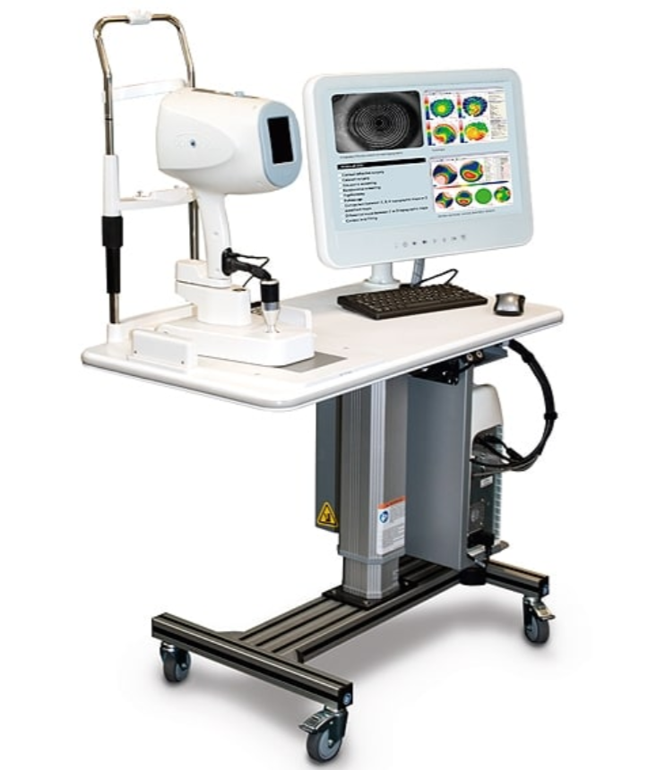

#SOCT Copernicus REVO with Angio OCT, Copernicus REVO is the new OCT Spectral Domain Optopol. It allows very high resolution scans in retina, optic disc and anterior segment miosis.
SOCT Copernicus REVO is very easy to use, which allows some personnel freedom. All you need to do is position the patient and start the unit. The automated examination process, which can be carried out even with minimal experience in the OCT domain, features a variety of analysis and review tools. This allows you to use it for either screening or diagnosis, depending on the patient’s needs and condition.
OCT made simple as never before
Position the patient and press the START button to acquire examinations of both eyes. The SOCT Copernicus REVO, using vocal messages, guides the patient through the process of increasing comfort and reducing patient chair time. Creating customized scanning protocols of different diagnostic scenarios will speed up workflow.
This OCT unit features a proprietary spectrometer that will guarantee faster scanning speeds and better quality images across a wider scanning surface. It will help you meet the daily demands of your practice, localize OCT services, and improve the patient experience. The ergonomically designed model also features noise reduction technology for a high definition presentation to improve detection and diagnosis capability.
SOCT Copernicus REVO Features:
Fully automatic. Alignment, focus, and acquisition are fully automated.
Extremely small dimensions and weights, like an auto refractometer.
Scans in mioses up to 12mm. Retina and disc are analyzed thanks to a single scan.
Front segment without any additional lens. It is possible to scan from both iridocorneal angles.
Connection to any PC thanks to a single USB cable.
Analysis of 8 retinal layers with the regulatory database.
Glaucoma analysis with nerve fiber thickness and Gangliari cell analysis with the regulatory database.
Pachymetric and iridocorneal angle analysis.
Methods of comparisons and follow-up of exams and pathologies.
Optional angiography module.
TECHNICAL DATA:
| Technology | Spectral Domain OCT |
| Light Source | SLED, Wavelength 840nm |
| Bandwidth | 50 nm half bandwidth |
| Scanning speed | 27000 A-scan per second |
| Axial resolution | 5 µm in tissue |
| Transverse Resolution | 12 µm, typical 18 µm |
| Overall scan depth | 2.4 mm |
| Scan range | 3 to 12 mm |
| Scan types | 3D, Radial, B-scan, Raster, Cross |
| Fundus image | Live Fundus Reconstruction |
| Alignment method | Fully automatic, Semi-automatic |
| Retina analysis | Retina thickness, Inner retinal thickness, Outer retinal thickness, RNFL+GCL+IPL thickness, GCL+IPL thickness, RNFL thickness, RPE deformation, IS/OS thickness |
| Glaucoma analysis | RNFL, ONH morphology, DDLS, Ganglion analysis as RNFL+GCL+IP and GCL+IPL, OU and Hemisphere asymmetry |
| Anterior | Pachymetry, LASIK flap, Angle Assessment, AIOP, AOD 500/750, TISA 500/750 |
| Anterior Wide Scan | Angle to Angle view, Adapter required |
| Min. pupil size | 3 mm |
| Focus adjustment range | -25D to +25D |
| Dimension/weight | 382 (W) x 549 (D) × 462 (H) mm/ 23Kg |
| Fixation target | OLED display (The target shape and position can be changed), External fixation arm |
| Power supply | 110-230 V, 60/50 Hz |

