Search for more information
μ-Scan Adaptive multi-ray imaging Dynamic color Single crystal transducers
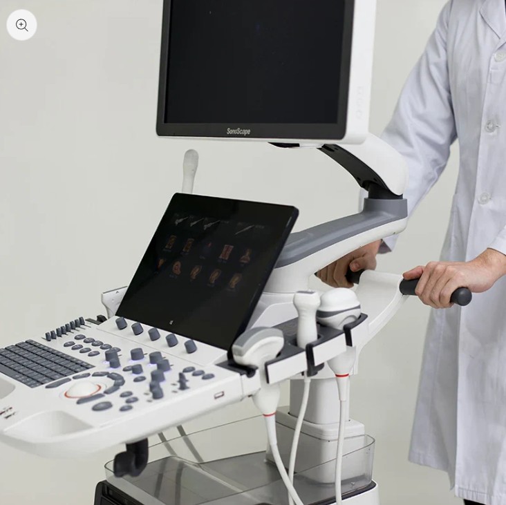
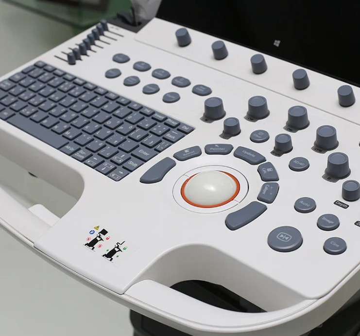
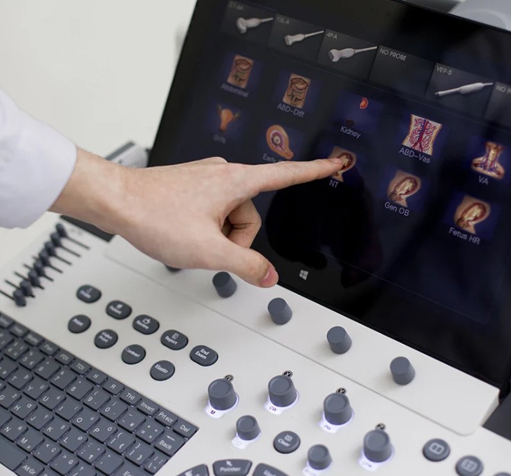
Ergonomic design 21.5″ LED monitor with articulated arm 13.3” adjustable touch screen Swivel and height adjustable control panel Integrated gel heater Wi-Fi connection
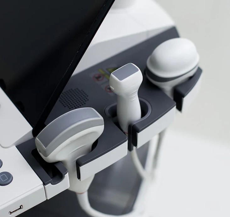
VC2-9 4D convex, 12LA linear, 6V3 vaginal
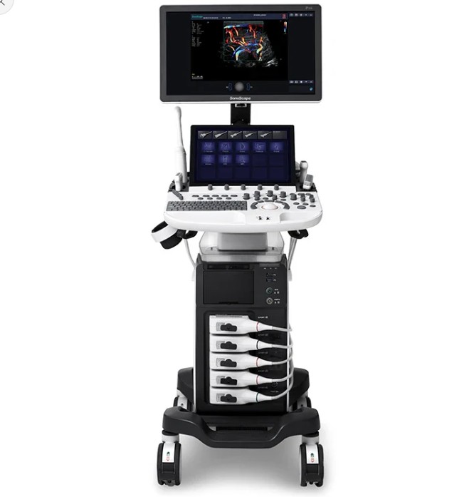
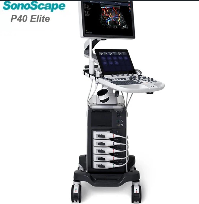
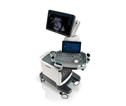
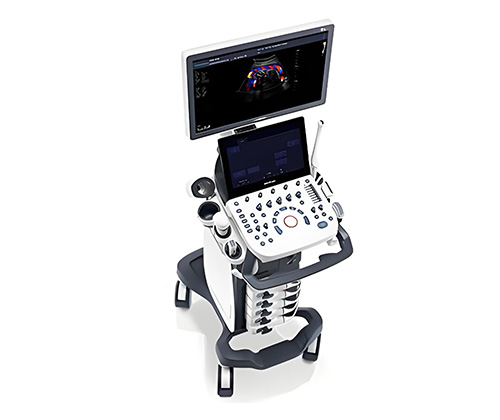
The SonoScape P40 Elite is a diagnostic ultrasound system designed for demanding clinical environments. It supports healthcare professionals in performing routine ultrasound imaging examinations. Designed to cope with heavy use, it promotes smooth organisation of the examination station. Its positioning is in line with a structured and precision-oriented medical practice.
Sonoscape P40 Elite ultrasound system
Specialties
Abdomen
Vascular
Cardiology
Gyn/OB
Urology
Musculoskeletal
Interventional ultrasound
Small parts
Anesthesiology
Pediatrics
Orthopedics
Cephalic
PF
Sonoscape P40 Ultrasound: Imaging Technologies
B/ 2B/ 4B/ M/ THI/ CFM/ PDI/ DirPDI/ PW/ HPRF/ CW
Dynamic Multi-beam technology
THI, PHI, TGC, LGC
μ-Scan
Auto (M-tuning)
Spatial Compound Imaging
Real-time 2D Panoramic
Color Doppler Panoramic
Widescan: Trapezoid Imaging
Freehand 3D Imaging
Auto NT
Auot EF
S-Guide
Auto IMT
Static 3D, 4D
S-Live (S-Live/ S-Live Silhouette)
Features
μ-Scan+: improved contrast and reduced artefacts in B and 3D/4D
SR-Flow: sensitive visualisation of low-speed flows
Micro F: microvascular flow analysis
S-Fetus: automation of obstetric planes and measurements
MFI Time: colour mapping of contrast-enhanced perfusion phases
Sono-synch: real-time sharing of the interface and camera for remote consultation

Advanced imaging architecture that leverages the new Single Crystal probes, the system expands diagnostic capability and quality in all clinical applications.
S-Thyroid is an advanced tool to detect and classify suspected thyroid lesions based on ACR TI-RADS (American College of Radiology Thyroid Imaging Reporting and Data System) guidelines. After selecting the region of interest, S-Thyroid can automatically define the boundaries of the lesion and generate a report on the characteristics of the suspected injury.
S-Brest is an advanced tool for detecting and classifying suspected breast injuries based on BI-RADS guidelines. After selecting the region of interest, S-Brest can automatically define the boundaries of the lesion and generate a report on the characteristics of the suspected injury.
S-Fetus is a user-friendly tool that allows fully automatic and accurate detection of the most significant plans and frequently used measurements of fetal biometrics. With a fetal head cine ring, S-Fetus can extract standard planes and display measurement results in a second, greatly reducing keystrokes and required working time by several times. It is designed to turn obstetric ultrasound into a much more welcoming, faster and enjoyable experience.
S-Pelvic is an advanced tool designed to reinvent the way doctors assess pelvic floor dysfunction (PFD). Thanks to highly intelligent capabilities, full automation of pelvic floor anatomy recognition, tracking and measurement are now available and can be achieved with a single click with unprecedented ease. In addition, S-Pelvic meets the 2D automatic front compartment rating and the 3D/4D automatic lever eltus rating and takes into account both Valsalva rest and maneuvering, with the aim of covering as many steps and details as possible in the pelvic floor ultrasound and offering a complete user experience.
Micro F provides an innovative method for expanding the visible flow range in ultrasound, especially for the visualization of small slow-flowing vessels. By adopting an advanced adaptive filter and accumulating temporal and spatial signals, Micro F can effectively distinguish minute flow from the movement of the overlapping tissue and represent hemodynamics with increased sensitivity and spatial resolution.
Bright Flow strengthens the definition of vessel boundaries by adding a 3D-like look to 2D color Doppler imaging. This innovative technology offers easy and improved spatial understanding and allows doctors to identify small blood flows as in a pop-off style. Luminous flux is also available for use in conjunction with other imaging modes, with the adjustable level of improvement, which offers more possibilities for clearer viewing.
Ergonomic design
21.5″ LED monitor with articulated arm
13.3” adjustable touch screen
Swivel and height adjustable control panel
Integrated gel heater
Wi-Fi connection

Compatible Accessories
Single Crystal C1-6 Probe
Single Crystal S1-5 Probe
12L-A Linear Probe
12L-B Linear Probe
9L-A Linear Probe
6V3 Probe
High-Performance Transducers
Single crystal transducers enable the pure imaging, especially for difficult patients, by increasing the uniformity of crystal alignment and raising energy transmission efficiency. Single Crystal C1-6 for abdominal and OB patients and S1-5 for cardiology and transcranial applications. Composite crystal linear transducers, by reforming the conventional piezoelectric materials, achieves a better acoustic spectrum and lower acoustic impedance to serve well in vascular, breast, thyroid, MSK, etc. The combo of 12L-a, 12L-B, 9L-A covers an ultra-wide frequency bandwidth, leaving nearly no blind spot for all sorts of scanning.