Search for more information

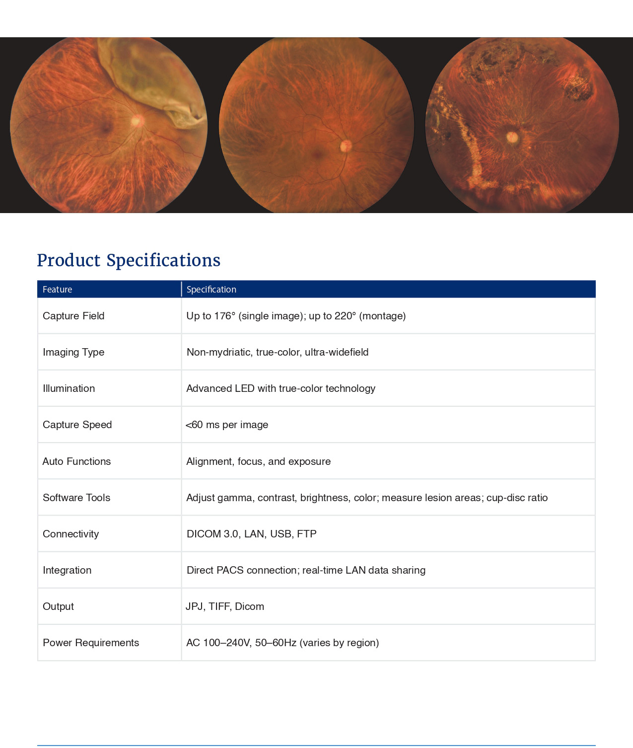
Wide field 176° (±5%) single shot Ultra wide field 220° (two images) Multi-mosaic > 260°
Approx. 12 megapixels (fundus image)
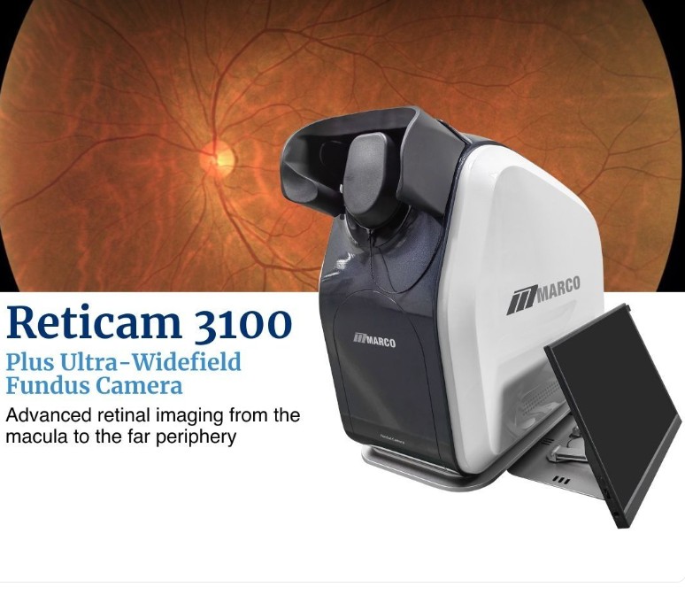
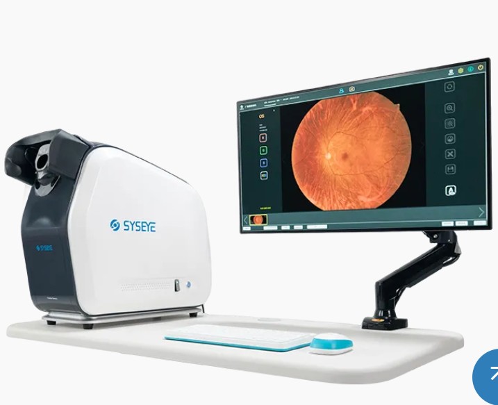
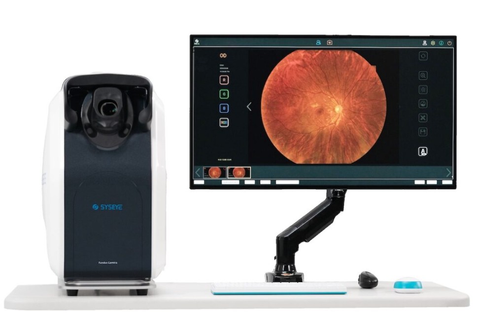
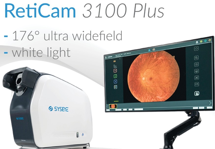
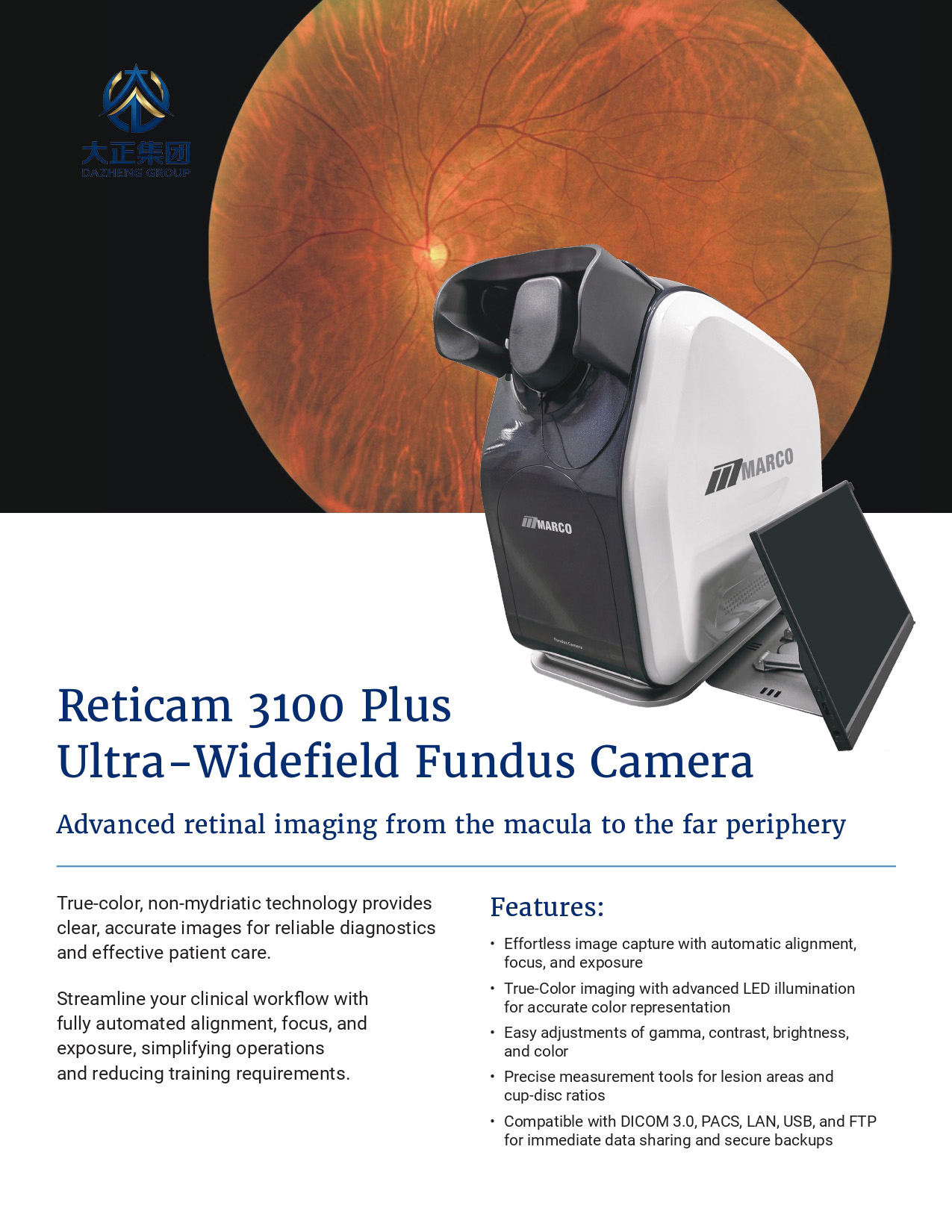
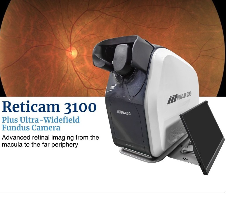
RetiCam 3100 Plus Ultra Wide-field Fundus Camera
Capture up to 176° in a single true-color fundus image and beyond 220° with automated mosaic
Supporting Retina Clinics, Screening Programs and Teaching Centers
With its combination of ultra-widefield coverage, true-color imaging, patient-friendly exposure, automated workflow and advanced software tools, RetiCam 3100 Plus is an excellent fit for:
Retina clinics managing diabetic retinopathy and macular edema
Hospital and community screening programs
Teaching centers and residency programs needing standardized images
Tele-ophthalmology and remote retina screening initiatives
Key Advantages
Have you ever imagined capturing more than 200° of the retina in a single, true-color view? With RetiCam 3100 Plus, automated mosaic technology makes it real — revealing retinal periphery that standard cameras simply leave out.
Up to 176° in one true-color image
Capture a 176° true-color fundus image from the eye center in a single shot, then automatically extend beyond 220° with mosaic — reducing the number of images needed and revealing more periphery.
LED soft exposure technology and maximum illumination <130,000 lx are designed to reduce pupil shrinkage and discomfort, making exams easier on children and light-sensitive patients.
Fast capture & high throughput
Ultra-fast acquisition lets you image many patients in a short time, helping clinics handle busy screening days and follow-up visits without compromising image quality.
Joystick-free, standardized imaging
Fully automated, joystick-free image acquisition — with automatic focus, exposure, alignment and working-distance adjustment — makes high-quality imaging repeatable across operators.
Offline AI & powerful review tools
Offline, unlimited AI assistance helps detect retinal changes without recurring cloud costs, while flexible software controls (brightness, contrast, gamma, RGB gain) make it easier to optimize and export images for training, teaching or AI development.
RetiCam 3100 Plus is designed for retina clinics, screening programs and teaching centers that need ultra-widefield coverage, true-color images, fast workflows and reliable, standardized image quality.
Real True Colors for Diagnosis & Teaching
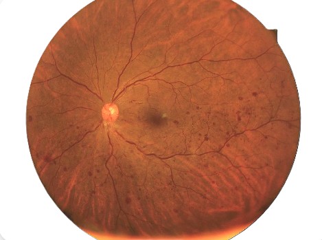
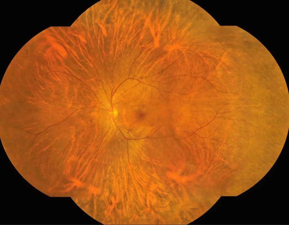
LED true-color imaging preserves natural fundus appearance, so physicians can interpret subtle changes with confidence and use images as a stable reference for teaching — without recalling patients just to confirm color accuracy.
Ultra-Wide Angle: Macula and Periphery Together
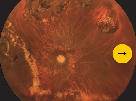
Up to 176° in a single shot and >220° with mosaic allow screening and diagnosis of macular disease (DR, AMD and more) while visualizing peripheral changes that narrow-field cameras may leave outside the frame.
Fully Automated, Joystick-Free Imaging
Automatic focus, exposure, alignment and working-distance control standardize image acquisition. Staff can be trained quickly, and consistent image quality becomes achievable regardless of operator experience.
Fully Automated, Joystick-Free Imaging
Automatic focus, exposure, alignment and working-distance control standardize image acquisition. Staff can be trained quickly, and consistent image quality becomes achievable regardless of operator experience.
Soft Exposure, Patient-Friendly Exams
Soft LED exposure and non-mydriatic operation are more comfortable for children and light-sensitive patients, helping to improve acceptance and cooperation during imaging.
Wide Angle Without Losing Resolution
RetiCam 3100 Plus maintains high optical resolution across its ultra wide field, so you see fine details not only at the posterior pole but also in mid-peripheral and peripheral retina.
Real True Colors for Diagnosis & Teaching
LED true-color imaging preserves natural fundus appearance, so physicians can interpret subtle changes with confidence and use images as a stable reference for teaching — without recalling patients just to confirm color accuracy.
Ultra-Wide Angle: Macula and Periphery Together
Up to 176° in a single shot and >220° with mosaic allow screening and diagnosis of macular disease (DR, AMD and more) while visualizing peripheral changes that narrow-field cameras may leave outside the frame.
Fully Automated, Joystick-Free Imaging
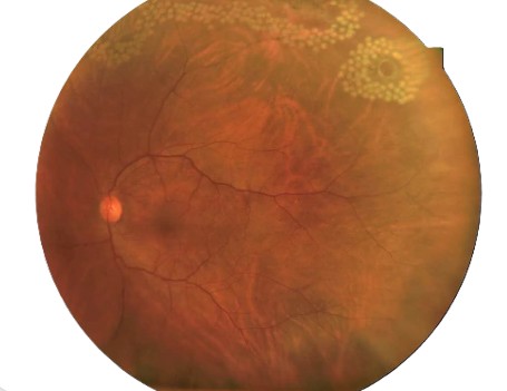
Automatic focus, exposure, alignment and working-distance control standardize image acquisition. Staff can be trained quickly, and consistent image quality becomes achievable regardless of operator experience.
High Throughput for Screening Workflows
Very fast image acquisition enables imaging many patients in a short time — ideal for community screening campaigns, satellite clinics and high-volume outpatient settings.