Search for more information
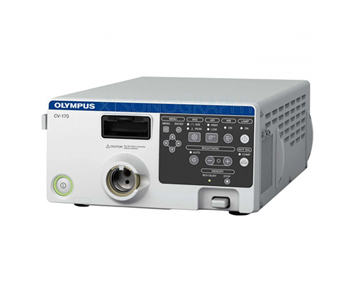
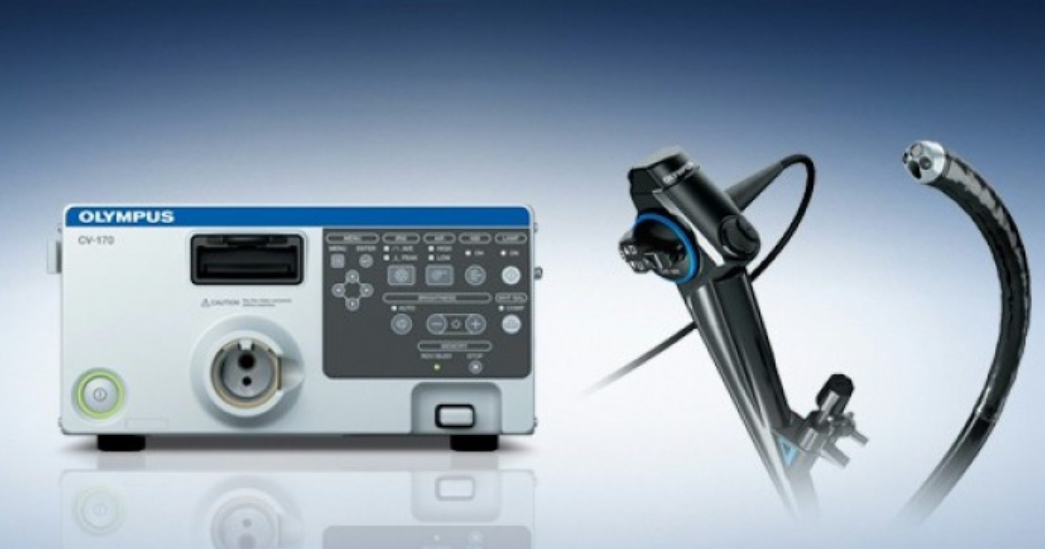
CV-170 – video endoscopic processor CV-170 Optera Power cable MAJ-2004 – keyboard MAJ-901 – water container (not used when working with video bronchoscopes) MH-155 – white cap MAJ-1925 – removable storage device MAJ-1951 – SDI cable Manual MAJ-1391 – foot switch
295 x 145 x 425 mm 11.0 kg
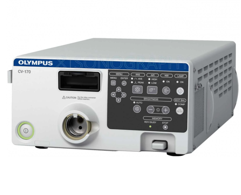
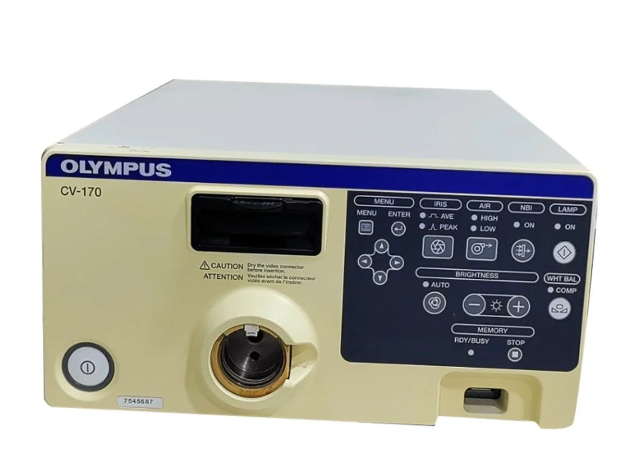
Olympus CV-170 Video Processor and LED Light Source
Discover the Olympus CV-170, an advanced video endoscopy system for enhanced medical diagnostics. With high-resolution imaging, dual-focus technology, and a user-friendly interface, it’s ideal for gastroenterology and ENT procedures. Embrace efficiency and accuracy in diagnostics with the compact and versatile CV-170.

Description
The Olympus CV-170 is an advanced video processor and endoscopy system designed for use in medical diagnostics and procedures. It is part of Olympus’ lineup of medical equipment, particularly for endoscopy.
Begin with an overview of the Olympus CV-170, emphasizing its role in modern endoscopic procedures. Highlight its compact design, efficiency, and advanced imaging capabilities, which make it a preferred choice in various medical environments.
Key Features of the Olympus CV-170
High-Quality Imaging: Explain how the CV-170 delivers clear, high-resolution images crucial for accurate diagnostics. Emphasize its capabilities in enhancing subtle features, which is important in identifying pathologies.
Dual Focus Technology: Discuss the system’s innovative dual focus technology, which allows for a detailed view of mucosal structures.
LED Light Source: Highlight the long-lasting LED light source, providing bright, consistent illumination while energy-efficient.
User-Friendly Interface: Mention its intuitive interface, allowing straightforward operation and efficient procedure workflows.
Narrow Band Imaging NBI: Thanks to the ability of hemoglobin to absorb light of a certain wavelength and scatter it in biological tissues, thereby contrasting blood vessels and mucosal structures, the CV-170 Optera is a powerful tool for the early diagnosis of cancer and focal mucosal lesions. Olympus uses two wavelengths, 415 nm (blue) and 540 nm (green), to maximize the photochromic function of hemoglobin.

Detail how the Olympus CV-170 is utilized in various medical procedures:
Gastroenterology: For endoscopic examinations like gastroscopies and colonoscopies.
Ear, Nose, and Throat (ENT): In ENT procedures, providing detailed views of the throat and nasal passages.
Advantages of Using the Olympus CV-170
Diagnostic Precision: Emphasize the system’s ability to provide clear and detailed images, leading to more accurate diagnoses.
Cost-Efficiency: The long-lasting LED light source reduces the need for frequent replacements and maintenance, making it a cost-effective choice.
Compact and Versatile: Its compact design suits various clinical environments, including smaller clinics and mobile medical units.
Ease of Use: Highlight how its user-friendly nature makes it accessible to a range of medical professionals.
Include:
CV-170 – video endoscopic processor CV-170 Optera
Power cable
MAJ-2004 – keyboard
MAJ-901 – water container (not used when working with video bronchoscopes)
MH-155 – white cap
MAJ-1925 – removable storage device
MAJ-1951 – SDI cable
Manual
MAJ-1391 – foot switch

Video System & Ancillary Devices
CV-170 All-in-One HD Video System w/LED & NBI
MAJ-1981 Olympus Keyboard
MAJ-1925 Olympus 2GB USB Portable Memory
MAJ-438 Remote Accessory Cable
MAJ-1981 Olympus Keyboard
MAJ-1925 Olympus 2GB USB Portable Memory
LMD-2110MD/OL 21.5″ LCD HD Widescreen Medical Grade Monitor
USB200 SD Image/Video Capture to USB Drive
IS40950 nStream DX V9.0 Image and Video Recording with DICOM
UP-25MD Sony Color Video Printer, small format (for office use)
TC-C2-PS Primary Endoscopy procedure cart with power supply and keyboard tray
AR-T10E Coupler
AR-T12E Coupler All-in-One HD Video System w/LED & NBI
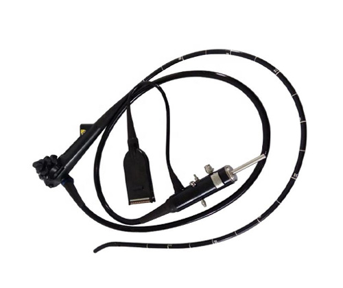
ENF-VH HD Rhinolaryngoscope

Product Specs
Power Supply
Voltage 100-240V AC: within +/- 10%
Frequency 50/60 Hz: within +/- 1 Hz
Rated Input 200 VA
Size
Dimensions (W x H x D) 295 x 145 x 425 mm (W x H x D)
Weight 11.0 kg
Observations
Examination Lamp LED Lamp
Analog HDTV signal output Either RGB (1080/60I: NTSC)/(1080/50I: PAL) or YPbPr (1080/60I: NTSC)/(1080/50I: PAL) output can be selected.
Analog SDTV signal output VBS composite (480/60I: NTSC)/(576/50I: PAL), Y/C (480/60I: NTSC)/(576/50I: PAL), and RGB (480/60I: NTSC)/(576/50I: PAL): simultaneous outputs possible.
Digital signal output HD-SDI (SMTPE 292M), SD-SDI (SMPTE 259M) and DVI (WUXGA, 1080p or SXGA) can be selected.
White balance adjustment White balance adjustment is possible using the white balance button on the front panel.
Color tone adjustments Red adjustment: ±8 steps, Blue adjustment: ±8 steps, Chroma adjustment: ±8 steps
Automatic gain control (AGC) The image can be electronically amplified when the light is inadequate due to the distal end of the endoscope being too far from the object.
Noise reduction Noise is corrected by image processing
Iris The auto iris modes can be selected using the “iris mode” switch on the front panel. Peak: The brightness is adjusted based on the brightest part of the endoscopic image. Average: the brightness is adjusted based on the average brightness of the endoscopic image.
Image enhancement setting Fine patterns or edges in the endoscopic images can be enhanced electrically to increase the image sharpness.
Either the structural enhancement or edge enhancement can be selected according to the user setup
Structural enhancement: Enhancement of contrast of the fine patterns in the image.
Edge enhancement: Enhancement of edges of the endoscopic image.
Freeze An endoscopic image is frozen using an endoscope or the “FREEZE” key on the keyboard
NBI Observation This is one of optical-digital observations using the narrow band observation light
Remote control DVR, Video printer, Image filing system, Flushing pump, Endoscopic CO2 regulation unit can be controlled (specified models only).
Documentation
Patient data Patient ID, Patient name, Sex, Age, Date of birth, Date of recording (time, stopwatch), Comments can be displayed in the endoscopic image screen.
Displaying the record state Portable memory and internal buffer, DVR, Video printer, Image filing system can be displayed on the monitor.
Advance registration of patient data Patient ID, Patient name, Sex and age, Date of birth of up to 50 patients can be registered.
Portable Memory
Media MAJ-1925 (OLYMPUS)
Recording Format TIFF: no compression, JPEG (1/5): approx. 1/5 compression, JPEG (1/10): approx. 1/10 compression
Number of recording images TIFF: approx. 227 images, JPEG (1/5): approx. 1024 images, JPEG (1/10): approx. 2048 images
Conclusion
Conclude by reinforcing the Olympus CV-170’s role in advancing endoscopic diagnostics, emphasizing its combination of high-quality imaging, cost-efficiency, and user-friendliness.