Search for more information
GE Vivid E95 GE Logiq E10 GE Logiq Totus GE Vivid S70 GE Logiq Fortis GE Logiq E9, S8, Voluson E8 Expert Vivid E9 systems.
Vascular, Breast, Small Parts, Musculoskeletal, Thyroid, Scrotal.


Product Overview
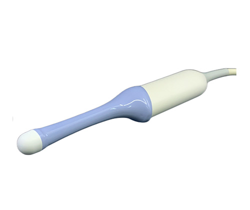
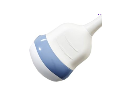
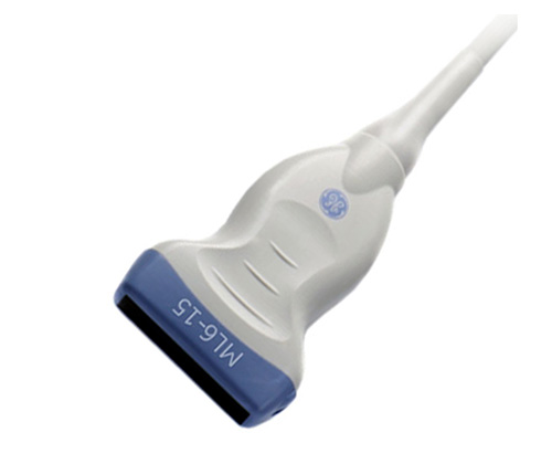
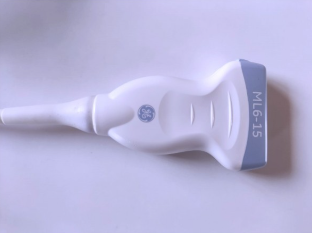
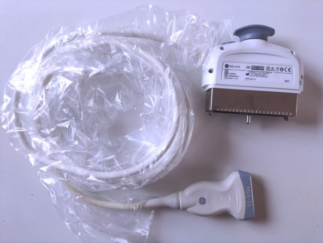
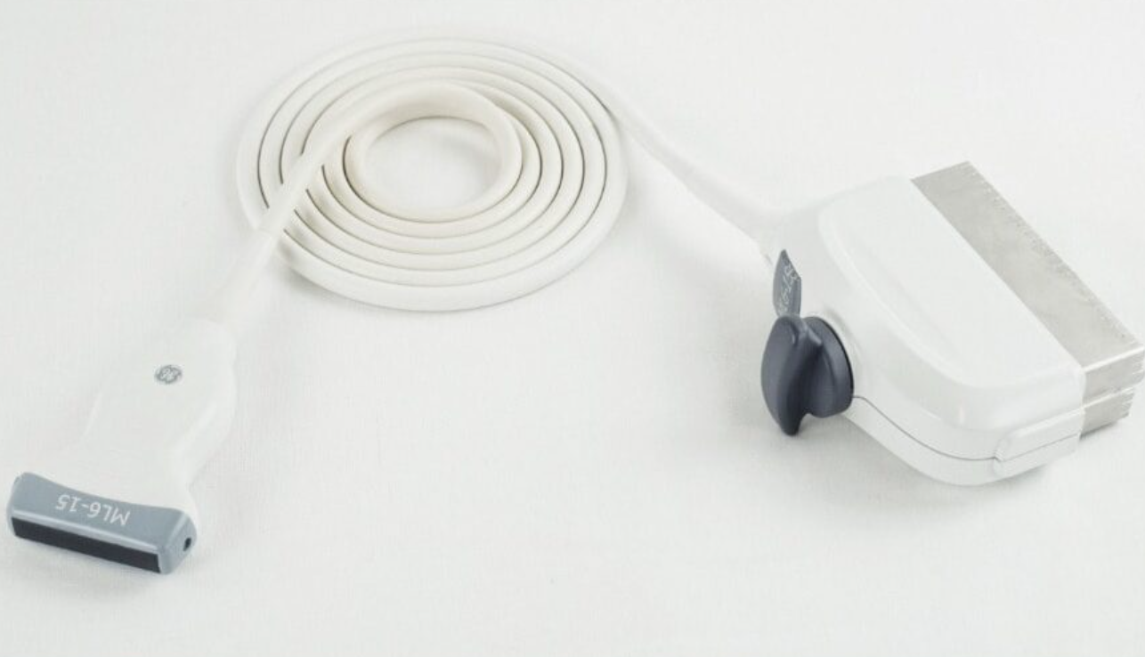
The ML6-15-D is a versatile ultrasound transducer designed to provide high-resolution imaging across a range of clinical applications. Its wideband matrix linear array technology ensures exceptional image quality, making it suitable for detailed examinations of small anatomical structures.
Applications: Vascular, Breast, Small Parts, Musculoskeletal, Thyroid, Scrotal.
GE Vivid E95
GE Logiq E10
GE Logiq Totus
GE Vivid S70
GE Logiq Fortis
GE Logiq E9, S8,
Voluson E8 Expert
Vivid E9 systems.
Technical Specifications
Frequency Range: 4 – 13 MHz, offering high-resolution imaging for superficial to mid-depth anatomical structures.
Scan Modes: B-Mode, Color Doppler, Pulse Wave Doppler.
Field of View: 49.6 mm aperture optimized for comprehensive visualization of target regions.
Depth of Field: Up to 6 cm, facilitating detailed imaging of superficial and mid-depth structures.
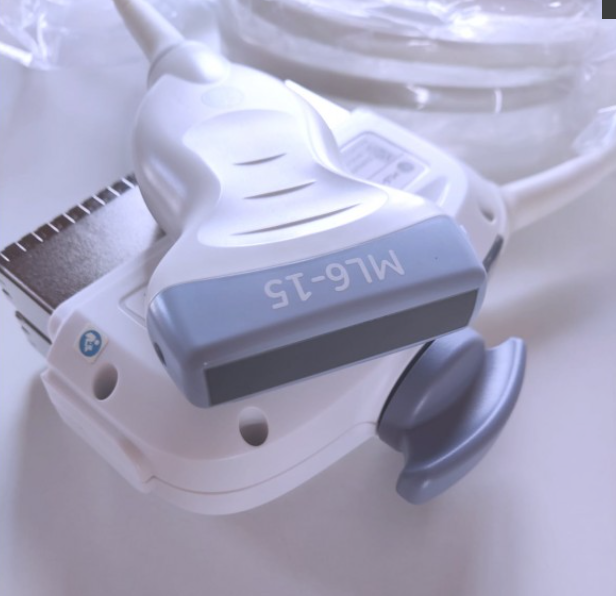
Footprint: Slim linear design enhancing access to anatomically challenging areas.
Connector Type: Standard GE proprietary connector, ensuring seamless integration with compatible ultrasound systems.
Design: Lightweight and ergonomic for improved operator comfort during extended use.
Applications
Small Parts Imaging: Provides detailed visualization of small anatomical structures such as the thyroid and breast.
Breast Imaging: Enables high-resolution assessment of breast tissue for diagnostic evaluations.
Musculoskeletal Imaging: Facilitates detailed examination of muscles, tendons, and ligaments.
Vascular Imaging: Supports accurate evaluation of vascular structures and blood flow dynamics.
Pediatric Imaging: Suitable for imaging small and delicate anatomical structures in pediatric patients.
Clinical Performance Highlights
High-frequency imaging ensures clear visualization of superficial anatomical structures, enhancing diagnostic confidence.
Advanced Doppler capabilities support precise assessment of vascular flow, crucial for vascular studies.
Reliable performance across a range of clinical applications, from routine examinations to specialized assessments.