Search for more information
Fast and efficient cell examination with integrated phase contrast (iPC) system Clear and wide visual field with 2X objective and FN 22 Experience 3D vision with inversion contrast (IVC) technology
Innovative Inverted Design High-Quality Imaging LED Illumination Phase Contrast and Fluorescence Capabilities
200mm×498mm×454mm 6.9KG
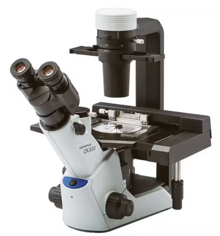

Explore the Olympus CKX53 Inverted Microscope, a breakthrough in cell culture observation and laboratory research. Offering high-quality imaging, LED illumination, and versatile capabilities for phase contrast and fluorescence, the CKX53 is ideal for biomedical research and clinical labs seeking detailed cell analysis and reliable performance.
Key Features of the Olympus CKX53
Innovative Inverted Design: Explain how the inverted design allows for easy observation of cell cultures in Petri dishes or flasks, a crucial aspect of cell biology research and clinical applications.
High-Quality Imaging: Highlight the microscope’s advanced optics that provide clear, high-contrast images, enabling detailed observation and documentation of cell cultures.
LED Illumination: Discuss the benefits of its energy-efficient, long-lasting LED light source, which provides consistent and optimal illumination for various samples.
Phase Contrast and Fluorescence Capabilities: Mention its phase contrast and fluorescence imaging options, essential for enhanced visualization of live cells and specific cellular components.

Why Labs Should Choose the Olympus CKX53
Enhanced Cell Observation: Emphasize the microscope’s ability to provide detailed views of cell cultures, aiding in accurate analysis and research.
User-Friendly Operation: The CKX53’s intuitive design and easy operation make it accessible to experienced researchers and beginners.
Versatility in Applications: Suitable for various applications, from basic cell culture to more advanced research involving live cell imaging and fluorescence.
Reliable and Durable: Built with Olympus’s quality standards, ensuring reliability and durability for long-term use in lab environments.

CKX53 Live cell microscopy
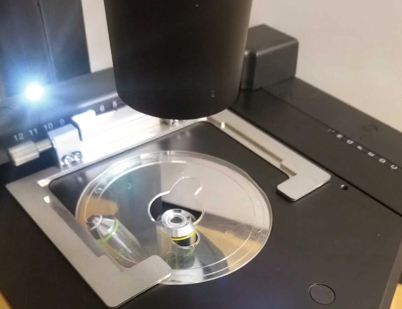
Fast and efficient cell examination with integrated phase contrast (iPC) system
Clear and wide visual field with 2X objective and FN 22
Experience 3D vision with inversion contrast (IVC) technology
User-oriented design for efficient cell sampling and easy handling
Efficient cell examination under sterile conditions
Simple cell sampling in sterile workbench conditions
Ergonomic, user-friendly design
Suitable for a wide range of cell culture containers
Better observation of multi-layer tissue culture flasks
Flexible container selection
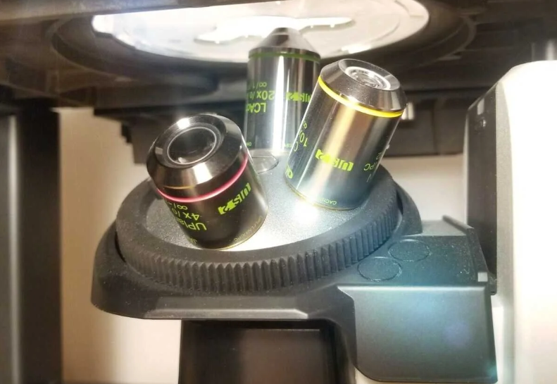
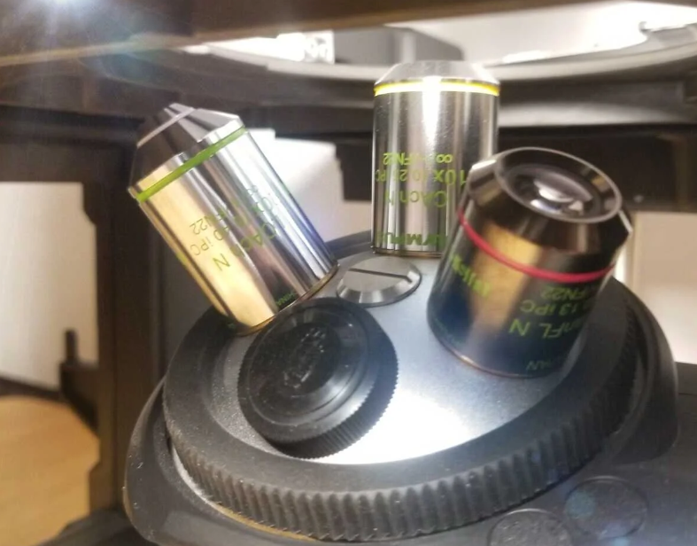
Efficient cell screening with a clear, wide field of view
The PLN2X objective aperture diaphragm slider has a 22mm field of view and a diameter of 11 mm
2X objective provides noticeably higher contrast than other objectives. Even transparent objects in the sample can be clearly identified
The wide field of view allows the detailed observation of individual cells in a 96-well plate without needing to move the stage

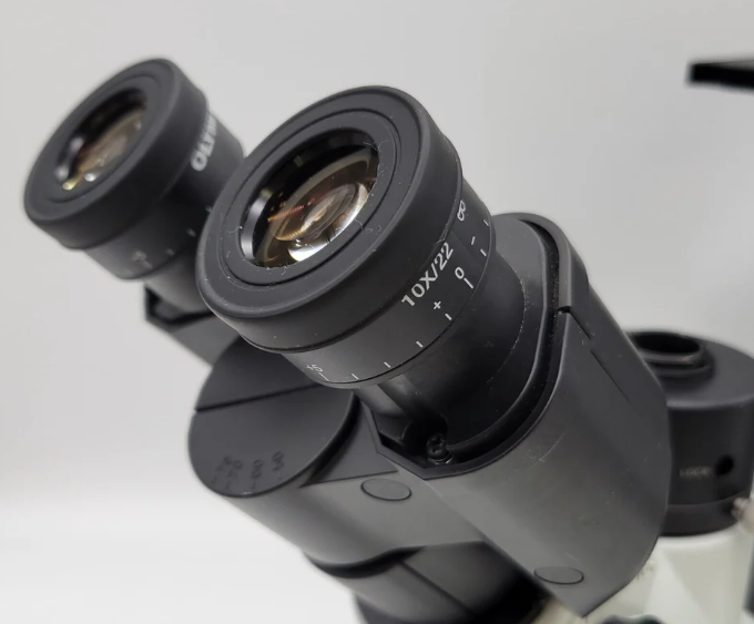
Comfortable viewing position
The 45-degree angle of the eyepieces and the positioning of the butterfly-shaped observation tube against the stage enables ergonomic cell observation, whether standing or seated.
Convenient hand position close to the controls
All the controls, including the power switch, coarse and fine focus, and the knob for switching the light path, are ergonomically arranged for ease of operation and reduced
With the CKX53 standard fluorescence set, even weak fluorescence signals can be viewed clearly thanks to a range of integrable light sources, such as a 100W mercury lamp (U-LH100HG), a 130W high-pressure mercury lamp (U-HGLGPS), as well as 3rd party LEDs (not available in all locations). The filter holder's three slots accommodate the same type of filter module as used with the IX3 and BX3 microscopes.
With the CKX53 standard fluorescence set, even weak fluorescence signals can be viewed clearly with the aid of different integrated light sources, such as a 100 W mercury lamp (U-LH100HG), a LED and LDP light source (U-LGPS), and 3rd party LEDs (Not available in some areas). The same type of mirror unit as those offered with our IX3 and BX3 microscopes can be set in the mirror unit slider’s three slots.
Further Specifications
Dimensions (W × D × H): 200 mm × 498 mm × 454 mm (phasecontrast standard configuration)
Weight: Approx. 6.9 kg