Search for more information
1,050nm wavelength
100,000 A-scans/second
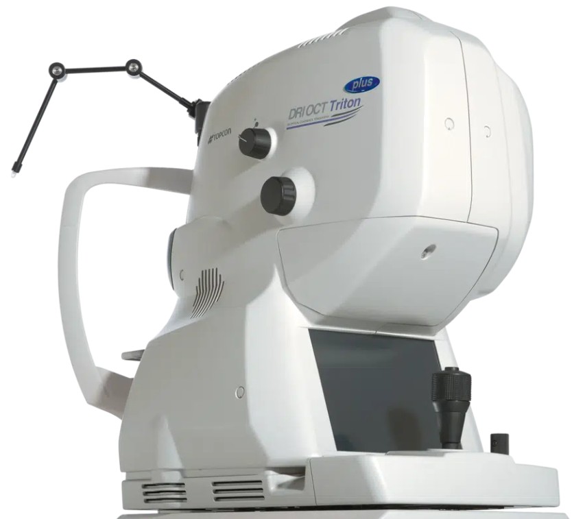
Topcon DRI OCT Triton Plus PC set with rack Electric optical stand Printer Anterior eye lens unit AA-1
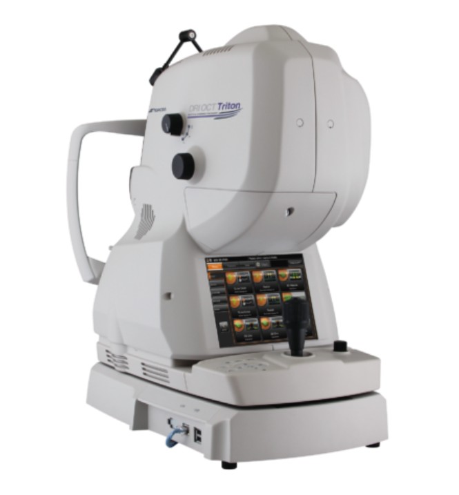
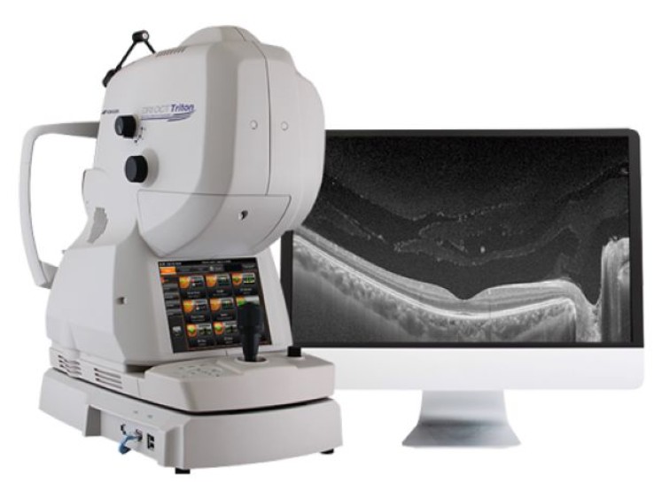
#Topcon DRI OCT Triton Swept source OCT——The DRI OCT Triton combines the world’s first Swept Source OCT technology with multimodal fundus imaging. Multimodal All-in-One fundus imaging tool will bring the next level of diagnostic capability to you and your patients
Combining comprehensive aspects of swept-source OCT, Topcon’s DRI OCT Triton ensures high-quality imaging. It takes into account various aspects of imaging to ensure high-resolution fundus imaging, which in turn guarantees better-quality images for more accurate assessments. These are some key features of the DRI OCT Triton that make it an ideal choice for your practice.

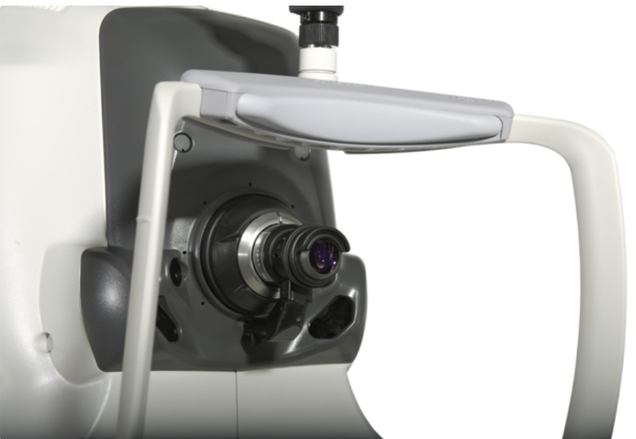
The Topcon DRI OCT Triton is a multimodal swept-source OCT system equipped with a non-mydriatic color fundus camera. Utilizing a 1,050 nm wavelength light source, and a scanning speed of 100,000 A Scans/sec, it provides uniform scanning sensitivity allowing superior visualization of the vitreous and choroid in the same scan.
Invisible OCT scanning light, eye tracking during the capture of selected scans, along with high scanning speeds reduce the effect of patient eye movement, improving workflow and allowing for more data be to collected in a shorter period. A 12 mm x 9 mm widefield scan along with automated layer segmentation provides measurement and topographical maps, with reference database, of the optic nerve and macula in one scan.
The Triton Plus model also includes a monochrome camera for fluorescein angiography and fundus autofluorescence utilizing the exclusive Spaide autofluorescence filters.
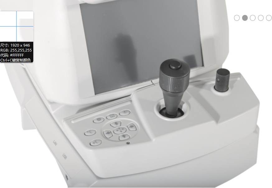
The easy-to-use, intuitive IMAGEnet 6 software enables dynamic viewing of the OCT data, providing 3D, 2D and fundus images simultaneously. Pin-Point™ Registration identifies an exact pathological location across all imaging modalities available within the Triton. In addition, both compare and trend analysis functions allow users to view serial exams of several scan protocols including our 12x9mm 3D Widefield scan. En face technology*, with layer flattening application allows for visualization of the various layers of the retina.
What does the DRI OCT Triton feature?
ncreased penetration
At 100,000A-Scans per second, which is roughly double the average scan speed, this OCT will offer better output than its competitors at an efficient rate. It features swept-source OCT, which is designed for improved visualization in a single scan. It also features a larger and deeper scan surface for increased detail.
Automatic tracking
The model features automatic scanning and tracking, which facilitate accurate imaging and monitoring of patients’ conditions. It automatically places any subsequent scans on specific positions to allow accurate monitoring of changes made from the initial scan results. This will enable a better assessment and facilitate higher-quality services for your patients. Active eye tracking will help map the eye, track any movement and account for the original structure to ensure that any blinks and sudden ocular movements do not compromise the accuracy of your findings.