Search for more information
Placido Disk Illumination: Led @635nm OCT Source: 5Led @845nm Pupillographic Illumination: Led @950nm
Placido Disk Rings: 22 Measured Points: 31232 (anterior surface) 25600 (posterior surface) Topographic Covering Range: 10 mm Dioptric Measurement Range: from 1D to 100D Measurement Accuracy: Class A according to UNI EN ISO 19980:2012
505 x 315 x 251 mm HxWxD 10.4 Kg
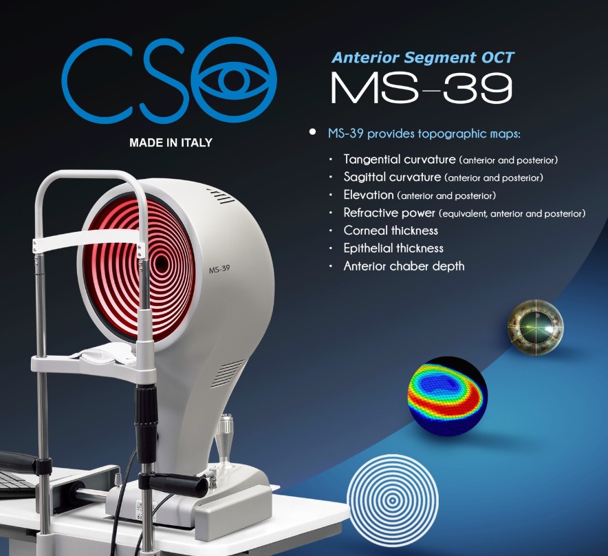
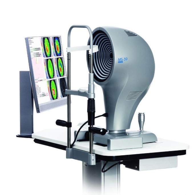
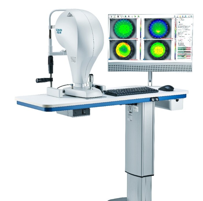
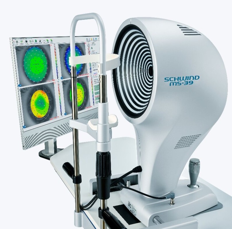
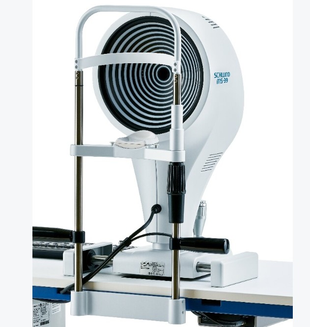
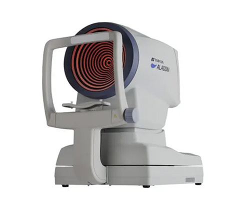
The AS-OCT MS-39 Corneal Topographer revolutionizes anterior eye imaging by combining corneal topography with high-resolution OCT anterior segment tomography. This advanced device provides exceptional clarity in cross-sectional images, offering precise details of the corneal structure. With its multiple features, it is essential for clinical eye diagnosis and corneal surgery planning.

More benefits
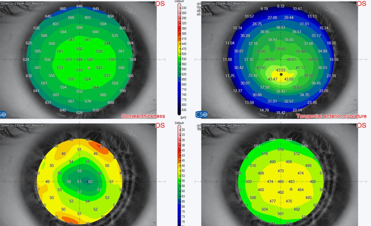
1、Keratoconus screening
This convenient tool provides important corneal data and can make a pre-operative contribution to avoiding possible complications associated with ectasia.
2、IOL calculation
The calculation module for intraocular lenses, based on ray tracing technology, is excellently suited for treatment planning of refractively treated or untreated eyes.
3、Intrastromal rings
Placement of intrastromal rings can be planned exactly with the help of pachymetry maps and a corneal elevation profile.
4、Glaucoma screening
Various useful screening parameters assist glaucoma specialists with diagnosis
5、Pupillography
The integrated function captures the pupil diameter dynamically or statically depending on the defined lighting conditions. Precise data concerning the pupil centre and diameter are essential for planning and performing most refractive treatments.
6、“Dry eye” analyses
The Placido technology allows for analysis of the condition of the tear film. This information provides support when diagnosing the symptoms of “dry eye”.
7、Lens biometrics
The crystalline lens thickness and their position in the eye can be precisely determined to support refinement of the IOL calculation

The MS-39AS-OCT epitomizes excellence in anterior eye analysis by merging Placido disk corneal topography with high-resolution OCT-based anterior segment tomography. This innovative fusion provides exceptionally clear cross-sectional images, revealing intricate details of the corneal structure and layers over a 16mm diameter. Beyond its diagnostic precision in the anterior segment, the MS-39AS-OCT also holds value in corneal surgical planning, particularly for refractive surgery. Additionally, a module for intraocular lens (IOL) power calculation, based on ray-tracing techniques, adds versatility to this invaluable tool for practitioners. Furthermore, with additional features such as precise pupil diameter measurements and in-depth tear film analysis, the MS-39AS-OCT offers a comprehensive solution for eye care professionals.
The MS-39AS-OCT fully leverages the Phoenix software, a robust platform that allows patient data to be stored for future review and analysis. This essential functionality ensures accurate patient tracking and facilitates information sharing across all CSO devices, thereby ensuring consistency in clinical data.
Keratoconus screening is a vital feature of the MS-39AS-OCT, providing clinicians with crucial insights into the patient's corneal health. By better understanding the corneal condition, practitioners can preempt potential complications associated with corneal ectasia before any surgical intervention.
Another notable feature of the MS-39AS-OCT is its ability to measure the epithelial layer advancedly, allowing for precise evaluation of any corneal surface anomalies. This detailed analysis contributes to more accurate diagnosis of ocular conditions, thereby enhancing the quality of care.
Aberrometric analysis offers a comprehensive view of corneal aberrations, essential for understanding or explaining patients' visual problems. Additionally, integrated pupillometry enables precise pupil diameter measurements in various lighting conditions, thereby contributing to optimizing visual quality.
The MS-39AS-OCT offers an intraocular lens (IOL) power calculation module based on ray-tracing techniques, enabling accurate calculation of spherical and toric power of the intraocular lens, regardless of the corneal condition.
Placido disk technology allows for advanced tear film analysis, including non-invasive tear film breakup time, essential for assessing ocular surface health. Moreover, crystalline biometry provides precise measurements of crystalline lens thickness, thereby contributing to more precise surgical planning.
Finally, the MS-39AS-OCT enables planning of intrastromal ring systems for correcting refractive defects and certain types of keratoconus. For glaucoma specialists, it offers the ability to measure anterior chamber angle parameters, corneal thickness, and useful values for disease diagnosis. With all these combined features, the MS-39AS-OCT represents an invaluable tool for eye care professionals, offering a detailed and precise analysis of the anterior eye.
Technical Details:
Data Transfer: USB 3.0
Power Supply: External power source 24 VDC, Input: 100-240Vac - 50/60Hz - 2A, Output: 24Vdc - 100W
Power Cable: IEC C14 plug
Dimensions (HxWxD): 505 x 315 x 251 mm
Weight: 10.4 kg
Chin Rest Movement: 70 mm ± 1 mm
Minimum Height of the Chin Cup from the Table: 233 mm
Base Movement (xyz): 105 x 110 x 30 mm
Working Distance: 74 mm
Light Sources:
Placido Disk Illumination: Led @635nm
OCT Source: 5Led @845nm
Pupillographic Illumination: Led @950nm
Topography:
Placido Disk Rings: 22
Measured Points: 31232 (anterior surface) 25600 (posterior surface)
Topographic Covering Range: 10 mm
Dioptric Measurement Range: from 1D to 100D
Measurement Accuracy: Class A according to UNI EN ISO 19980:2012
Section:
Image Field: 16 mm x 8 mm
Axial Resolution: 3.6 µm (in tissue)
Transversal Resolution: 35 µm (in air)
Image(s) Resolution: Keratoscopy (640x480) + 25 radial scans on a 16 mm transversal field (1024 A-scan), Section: 1.6 mm (1600 A-scan) on 8 mm (800 A-scan)
Operating System: Windows 10 (64 bits)
Features:
Combination of corneal topography and anterior segment tomography by OCT
Provides clear and detailed cross-sectional images
Precise measurements of pachymetry, elevation, curvature, and dioptric power of the cornea
Intraocular lens calculation module based on ray-tracing technique
Advanced analysis of tear film with Placido disk technology
Precise measurement of pupil diameter with integrated pupillometry