Search for more information
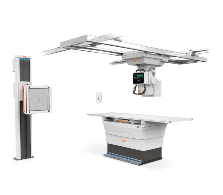
CARESTREAM DRX-Evolution Plus System
The Carestream DRX-Evolution Plus offers:
• New LED lighting for enhanced functionality and aesthetics
• Greater flexibility with an extended tube column
• High-performance Carestream generator
• Optional table to accommodate patients up to 705 lbs (320 kg)
• Forward-looking design to accommodate advanced imaging applications in the future
Choose a detector that best fits your imaging needs:
DRX Plus 3543 and 4343 for general radiology.
DRX Plus 3543C and 4343C for pediatrics and other dose-sensitive applications.
DRX 2530C – for dose-sensitive pediatric applications and orthopaedic imaging.
Carestream’s three DRX detectors offer:
Wireless connectivity to eliminate the hassles and hazards of cables and reduce the risk of infection.
Instant access to images to speed decisions on patient care.
Ability to share detectors between any DRX room or mobile DRX system.
A separate battery charger that allows the detector to be used while a spare battery is being charged.
Proven Image Quality
The wireless, cassette-sized DRX Detector and EVP Plus image-processing software deliver superb image quality.
Optional EVP Plus Software provides increased latitude and high-contrast image detail.
Software options accommodate specialities such as paediatrics, orthopaedics, and trauma.
Versatility
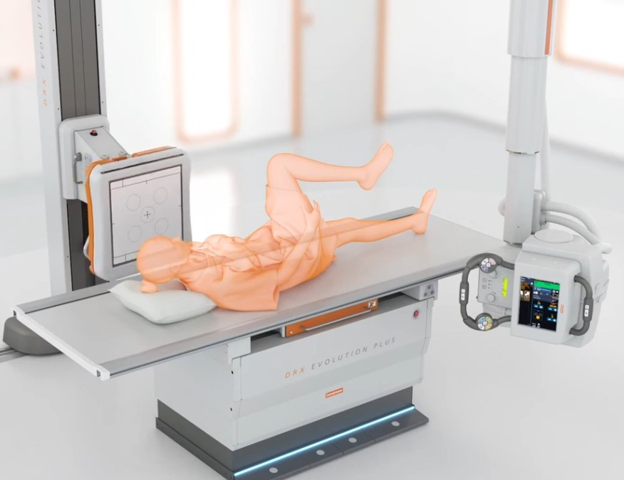
Choose a one-, two- or three-detector option to perform many types of imaging exams, including tabletop and long-length imaging.
The automated overhead tube is designed for positioning convenience and efficiency.
Detectors are fixed or wireless.
Scalability
Customize a DR solution to fits your workflow and budget
Add DR systems as your facility expands and grows
Designed for advanced future applications
Increased Productivity
Images are ready for viewing and manipulation on the console and in seconds
One space-saving console controls all medical imaging functions
Auto-positioning for fast and accurate setup
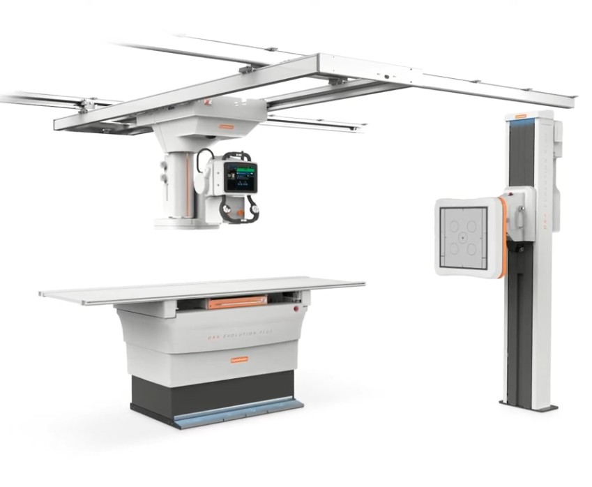
COMPONENTS
DRX Detectors
DRX-1 Detector with gadolinium (GOS) scintillator
DRX-1C Detector with cesium iodide (Csl), with increased DQE and MTF
DRX 2530C Detector with a smaller format design
A 17 in. x 17 in. (43 x 43 cm) stationary GOS detector, or a low-dose, cesium (CsI) panel option
Table Options
Standard table for patients up to 600 lbs. (272 kg)
High weight capacity for patients up to 705 lbs. (320 kg.)
Both feature a four-way floating top
Table movement initiated through double-tap foot pedals
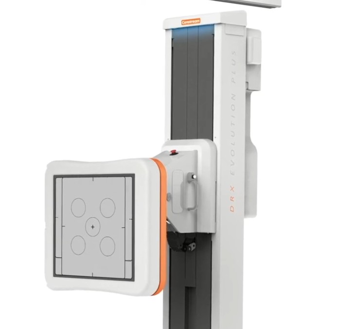
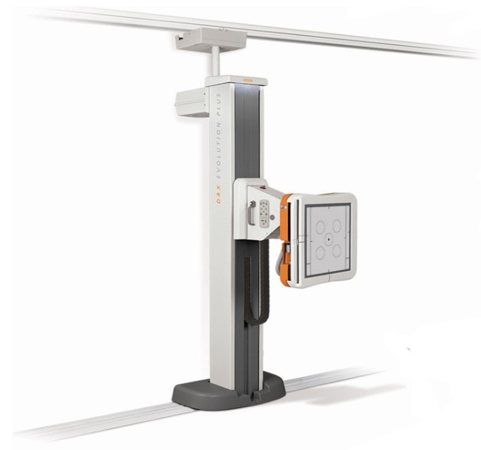
Motorized Wall Stand
Vertical Bucky range of motion from the floor up to 71 in. (180 cm)
Horizontal projections with tilt capability from negative vertical (–20°) to horizontal (90°)
Optional floor-mounted rail system for lateral stand movement
Auto tracking and centring align detector with overhead tube
A broad range of studies – chest, lateral cervical spine, standing knee and more
Horizontal Bucky positioning for upper extremity and under-the-table studies
Side-to-side swing angulations for easy cross-table exams on gurneys
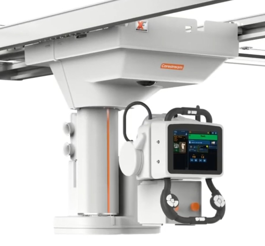
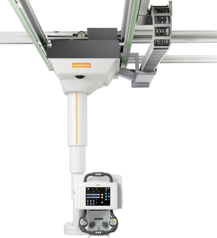
Motorized Overhead Tube
Preprogrammed tube and Bucky positions, generator and collimator settings maximize efficiency
Auto-positioning and auto-centring for table or wall stand procedures
Auto-tracking directs the tube to follow the detector to correct location or move the tube and the detector will follow
The asymmetric-collimation capability allows top or bottom blade to remain fixed while moving the other
Motor-assist function provides easy manual overhead tube positioning reducing operator fatigue
Non-Motorized Overhead Tube
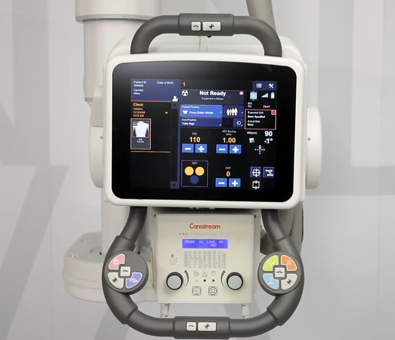
Monitor exams accurately with colour LCD’s self-righting display of techniques and key data including SID, kVp/mAs, exam type, tube-head angle, active detector, auto-centring and auto-tracking
Simple collimator controls positioned for easy adjustment
Automatic collimator option with additional internal filtration available
Operator Console
The flexible user interface, with touch screen control, customized to match the clinical workflow
Supports Automated Procedure Recognition (APR)
Supports workflow protocols such as DxIOD, IHE Scheduled Workflow, IHE Consistent Presentation of Images and IHE Dose Reporting