Search for more information
110V‐240VAC 50/60 Hz 330 mm x 220 mm x 320 mm 7.1Kg
Speed is adjustable; Sound can be played back.
4 Segments
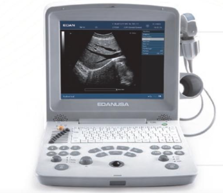
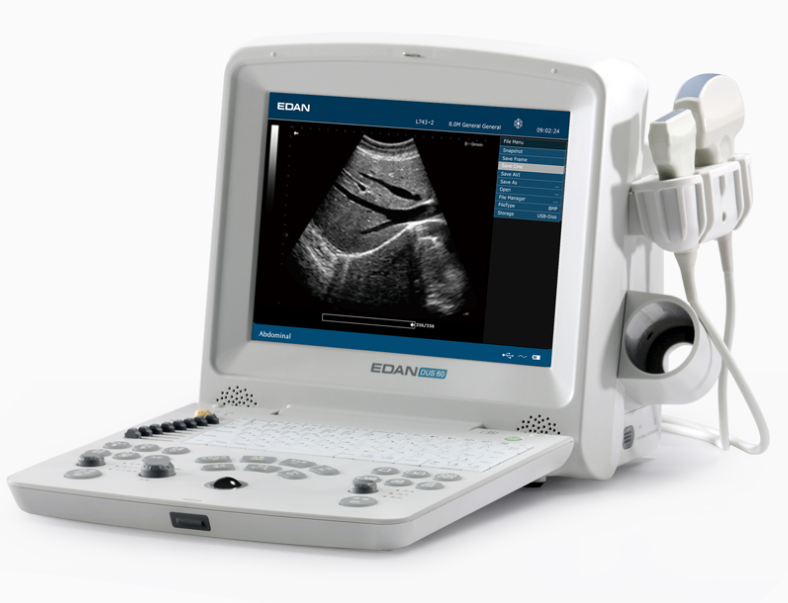
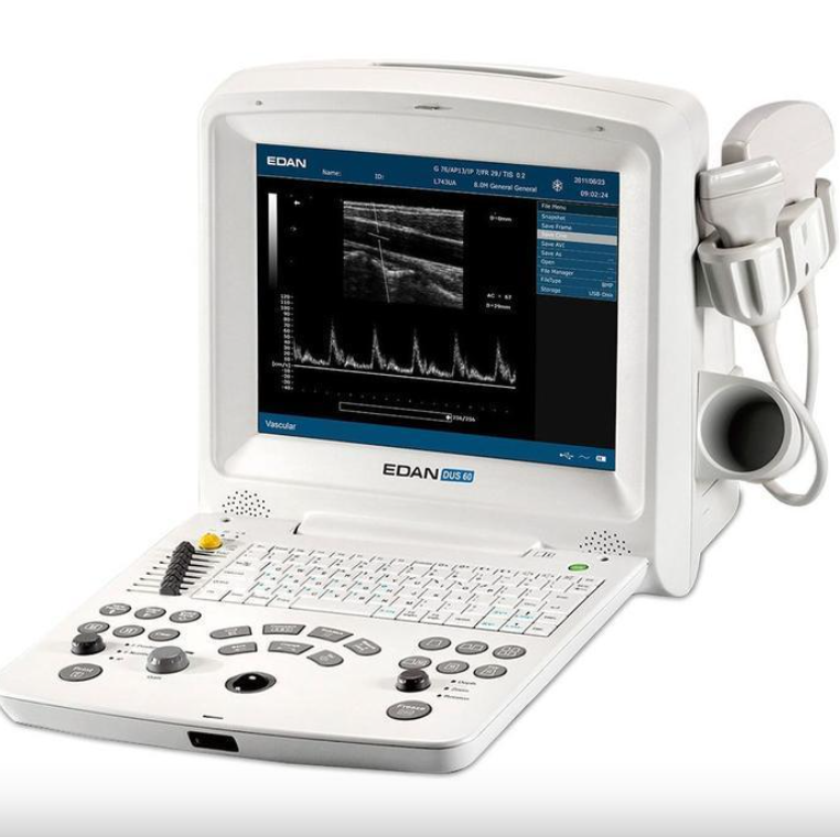
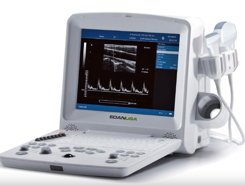
The EDAN DUS 60 is an impressive new compact ultrasound system, providing superb value across a large range of applications with its PW Doppler capabilities.

Powerful Technologies to Increase Your Diagnostic Confidence:
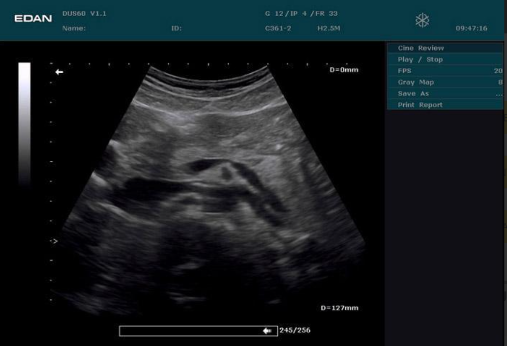
-Phase Inversion Harmonic Imaging technology provides best-in-class image quality.
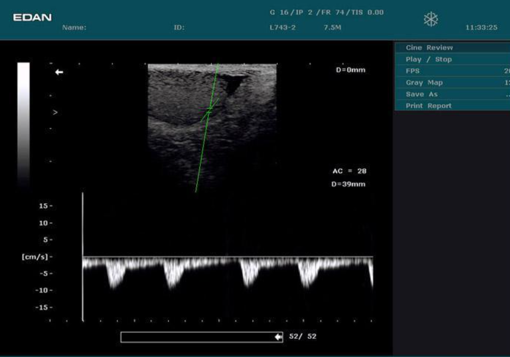
-PW Doppler supplies physiologic information for increased diagnostic value.
-Multiple transducer options increase versatility.
Go Anywhere You Need to Go:
-Compact and lightweight design for excellent mobility.
-Built-in battery provides up to 2 hours of point-of-care imaging.
-Large capacity data storage (504 MB).
Intuitive User-Friendly Design:
-One touch image optimization via smart IP key.
-Backlit, easy-to-use control panel.
-User-defined keys to customize your work-flow.

Practical Tools Enhance Efficiency:
-Intelligent 8-segment TGC for precise adjustment.
-Multi-format data transfer via USB and DICOM 3.0 (optional).
-7 pseudo-color options enhance image presentation.
Edan DUS 60 Digital Ultrasonic Diagnostic Imaging System Specifications:
General:
-Gray Scales: 256.
-Scanning Angle: Up to 152° (transducer dependent).
-Scanning Depth: From 19 mm to 324 mm (transducer dependent).
Functions:
-Body Mark: > 130 types.
-Transducer auto-detection.
Peripheral Ports:
S-video output, Video output, VGA output, 2 USB ports, Ethernet port, Remote control, and Footswitch port.
Measurement and Calculation:
B-mode: Distance, circumference, area, volume, angle, ratio, %stenosis, histogram.
M-mode: Distance, time, heart rate, slope.
Doppler: Time, heart rate, velocity, acceleration, trace, and RI.
More EDAN DUS 60 Ultrasound System Specifications:
Power Supply: 100 - 240 V (50/60 Hz).
Dimensions: 13.0" L x 8.7" W x 12.6" H (330 mm x 220 mm x 320 mm).
Net Weight: 15.7 lbs (7.1 kg).

Applied Technologies:
Tissue Specific Imaging (TSI), Tissue Harmonic Imaging (THI), Digital Beam-Forming (DBF), Dynamic Receiving Focusing (DRF), Real-time Dynamic Aperture (RDA), Dynamic Frequency Scanning (DFS), and Dynamic Apodization.
Display Modes: B, B+B, 4B, B+M, M, and PW.
Display: 12.1" TFT-LCD monitor with date, Time, Probe Name, Probe Frequency, Frame Rate, Patient Name, Patient ID, Hospital Name, Depth, Exam Type, Measurement Values, Body Marks, Annotations, and Probe Position.
Transducer Connections: 2 standard transducer connectors with a maximum frequency up to 10 MHz.