Search for more information
Multi area specular microscopy Enhanced usability and quick analysis Advanced manual analysis functions Combination of auto and manual analyses Additional features with CEM Viewer for NAVIS-EX
New
In Stock
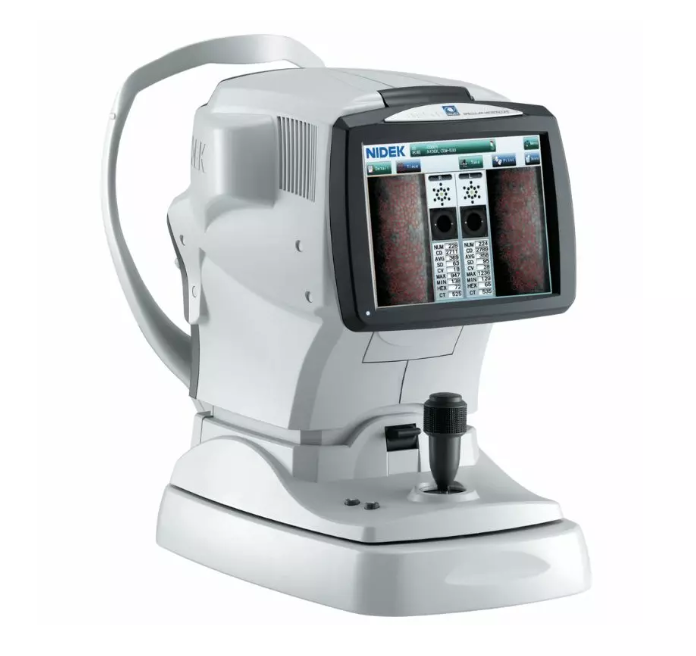
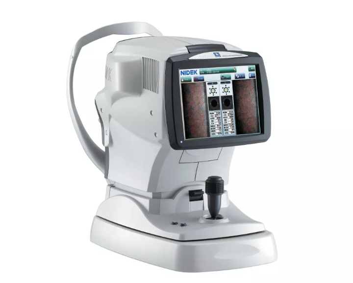
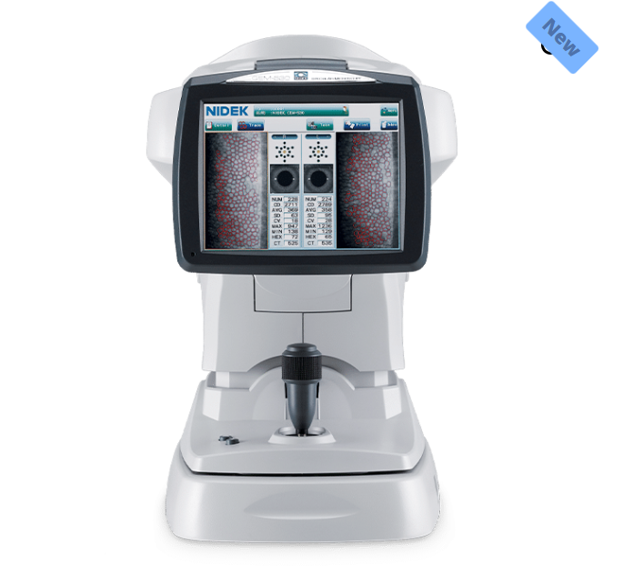
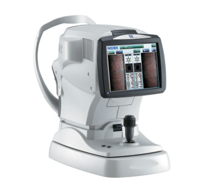
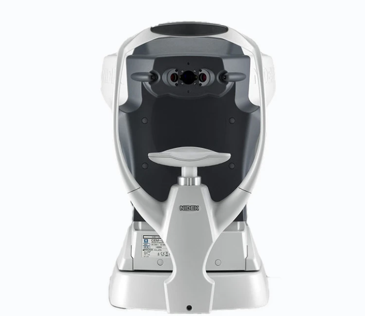
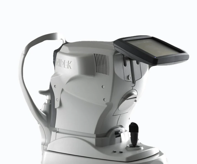
#CEM-530 Specular Microscope
A corneal endothelial examination is an optical test used to examine the cornea, specifically the corneal endothelial cells, which maintain corneal transparency. Because the number, shape, and size of corneal endothelial cells are not informative, a corneal endothelial examination is mandatory for all patients undergoing cataract surgery.
Unique Built-in Function: Paracentral Mode
The combination of central, paracentral, and peripheral imaging provides a broader, overall view that can be used for detailed morphological and quantitative evaluation of the endothelial layer and individual cells.
Seamless Imaging from Start to Finish
With 3D auto tracking and auto shot, vision practitioners can now easily choose optimal images for analyzing data, get comprehensive analysis in 2 seconds, and view endothelial cells in 4 different modes.
Enhance CEM-530 with CEM Viewer for NAVIS-EX
An optional function vision practitioners can choose is the CEM Viewer – a software for viewing and working with data using NAVIS-EX. Features include data management and endothelial cell count, progression follow-up and comparison, and paracentral display with peripheral.
Multi-Area Specular Microscopy
The combination of central, paracentral (8 points), and peripheral (6 points) imaging provides a broader, overall view that can be used for detailed morphological and quantitative evaluation of the endothelial layer and individual cells.

Seamless Auto & Manual Integrations
Rapid analysis increases the efficiency of the practice. Once the image is selected, complete analysis is automatically performed in two seconds with the CEM-530.
The analysis screen allows visualization of the endothelial cells in four modes, trace, photo, area, and apex, which helps the clinician to verify analysis values with the correspondent cell images.
2 Histograms
In Shape (Pleomorphism) and Size (Polymegathism)¹
2 Seconds
Auto Analysis with 4 Modes for Viewing Endothelial Cells
The CEM-530 provides comprehensive analysis including two histograms of variation in shape (pleomorphism) and size (polymegathism). For detailed analysis, the range of analysis can be changed and the cells to be excluded can be selected at the user’s discretion.

Specifications
| Device | Specular Microscope |
| Model | CEM-530 |
| Type | Diagnostic |
| Specialty | Cornea |
| Capture Field | 0.25 (W) x 0.55 (H) mm |
| Capture Position | Central 1 point Paracentral 8 points (5º Visual Angle, 45º Spacing) Peripheral 6 points (27º Visual Angle, 60º Spacing) |
| Measurement Range | 300 to 1,000 μm Accuracy ±10 μm |
| Auto Tracking | X-Y-Z Direction |
| Auto Shot | 16 Images Automatically Captured Indicates Optimal Image for Analysis |
| 4 Viewing Modes | Trace, Photo, Area, Apex |
| 2 Variated Histograms | Shape (Pleomorphism) Size (Polymegathism) |
| Display | Tiltable 8.4″ Color LCD Touch Screen |
| Ports | LAN, USB, Video Output Accessory BNC connector for video printer |
| Dimensions | Width: 11.5 in (291 mm) Depth: 19.5 in (495 mm) Height: 18.0 in (457mm) |
| Weight | 20 Kg |
| Standard | Built-In Thermal Line Printer |
| Optional Add-Ons | External Video Printer CEM Viewer for NAVIS-EX |