Search for more information
2D Imaging Tissue Harmonic Imaging M-Mode Velocity Color Doppler Color Power Doppler PW PW Tissue Doppler CW Doppler angle Correct after freeze
32.6 cm x 30.7 cm x 6.4 cm (L x W x H) 4.18 kg
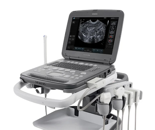
#The refurbished Sonosite Edge II is an upgrade from the original Sonosite Edge with new technology such as DirectClear. This ultrasound machine is very durable and provides the user with great image quality.
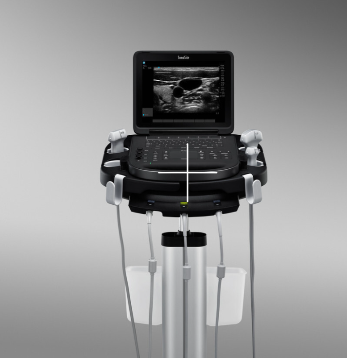
VISUALIZATION, CLEARLY ENHANCED
In the acute care environment, reducing the time to make an accurate diagnosis is a critical need. The Edge II features DirectClear Technology, a novel, patent-pending process that is available on select transducers. DirectClear elevates transducer performance by increasing penetration and contrast resolution. This contributes to an unsurpassed imaging experience for the bedside clinician.

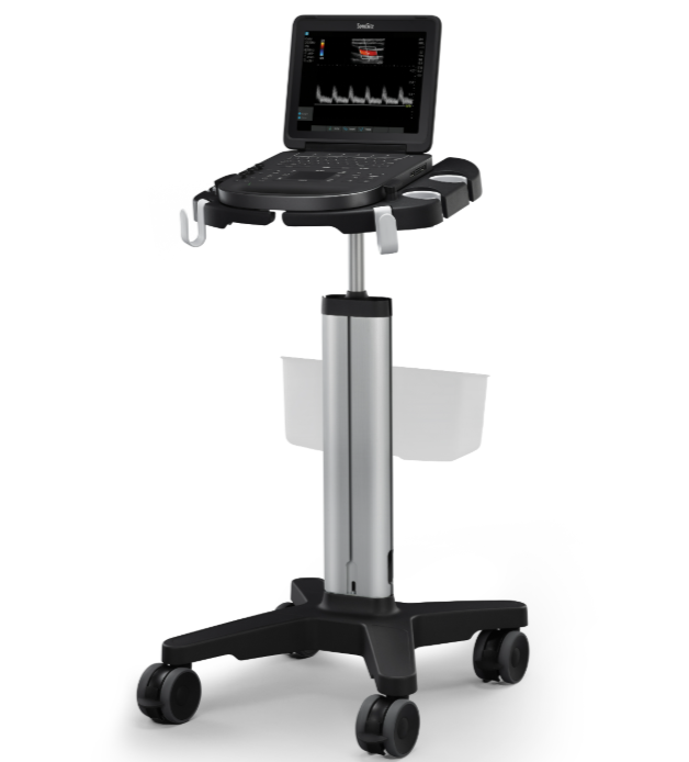
Portable, suitable for use with or without the optional Sonosite Edge Stand
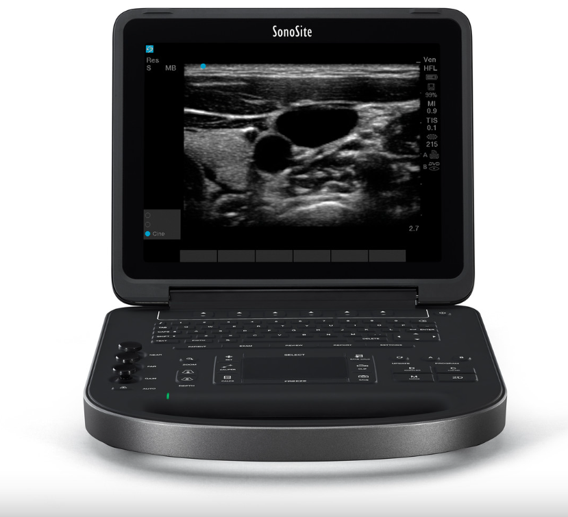
Wide-angle display featuring an anti-reflection coating ensures a clear view under varying lighting conditions
Streamlined interface for easy access to commonly used features. The trackpad, coupled with a select button, facilitates smooth navigation
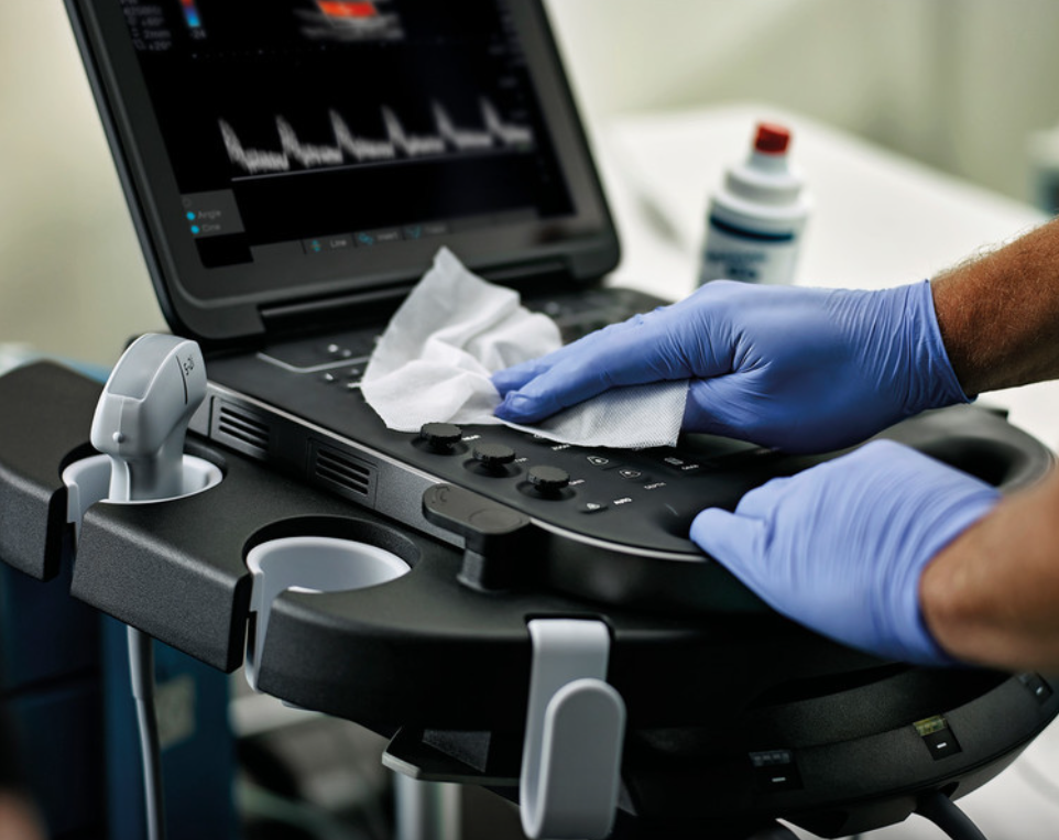
Low-profile buttons utilize snap dome technology, making them easy to clean and providing tactile feedback
The silicone keypad is seamlessly sealed to the edge, preventing liquid ingress and enabling effortless disinfection
Rapid cold start scanning readiness in under 25 seconds
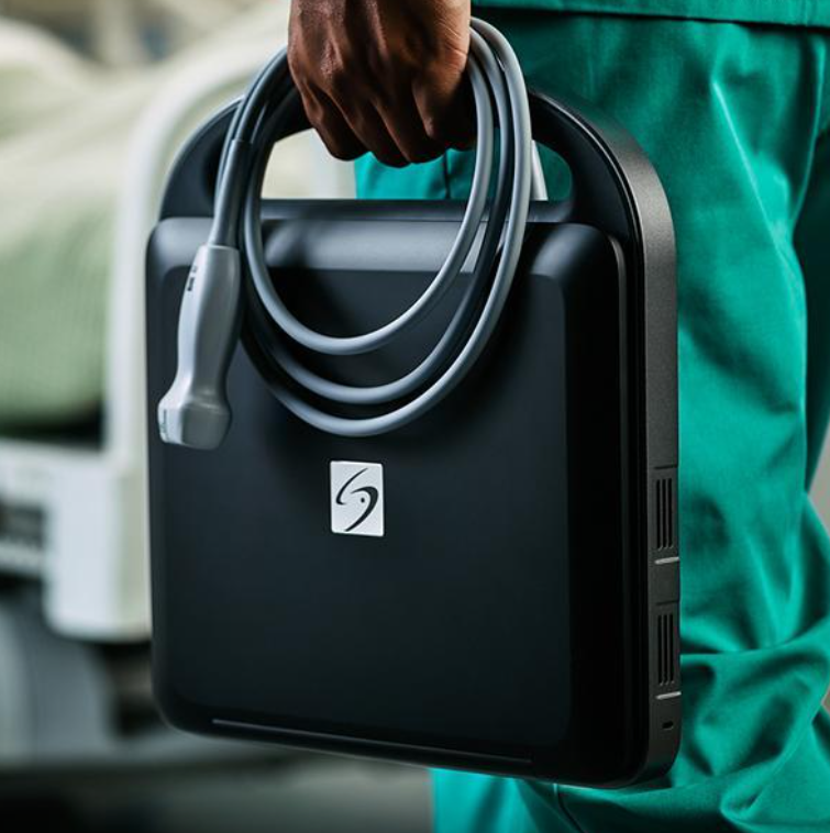
Optional Armored Cable Technology shields transducer cables from potential accidental damage
TAKING DURABILITY TO THE ARMORED LEVEL
Designed to be truly portable and used in the most rugged environments, the Edge II reinforces our commitment to reliability and durability with Armored Cable Technology. With an embedded metal jacket, armored cables protect transducers from common abuse accident scenarios, like being rolled over, stepped on, or twisted, and help maintain image quality over the life of transducers built on this cable platform.
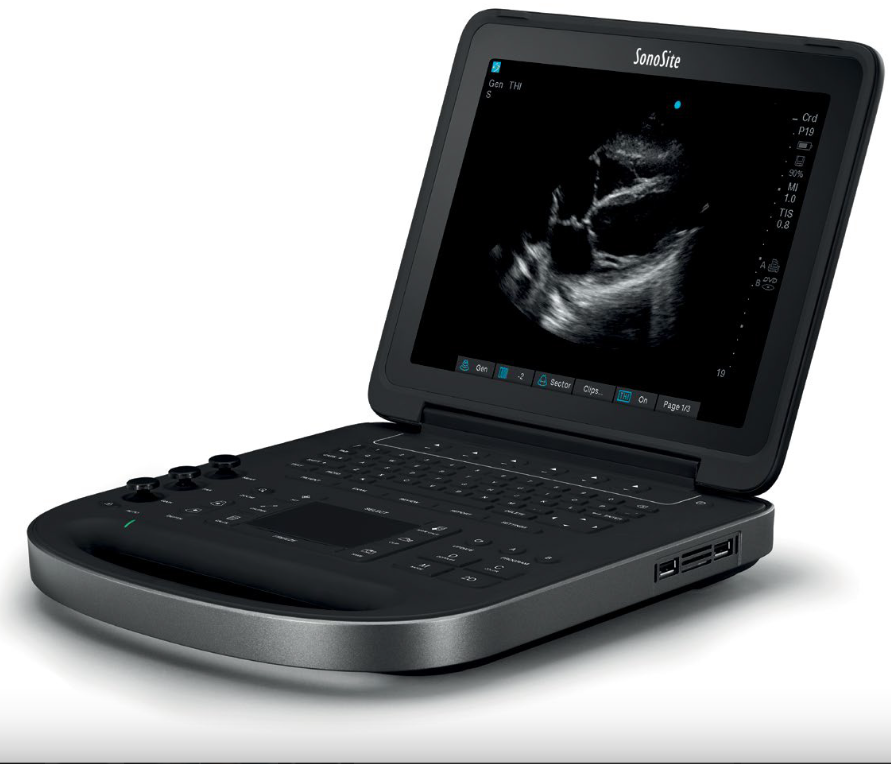
EDGE II ULTRASOUND FOR CLARITY AND CONFIDENCE
The Edge II features an easy-to-use interface for intuitive access to frequently used functions like gain and a wide-angle display with an anti-reflection coating for minimal adjustments during viewing. Designed with infection control in mind, the Edge II boasts a keypad with a seal extended to the edge to inhibit liquid ingress and low-profile keys with snap-dome technology for easy cleaning and tactile feedback.

SONSITE EDGE II SYSTEM SPECIFICATIONS
System Weight: 9.21 lbs/4.18 kg with battery
Dimensions: 12.8" x 12.1" x 2.5"/ 32.6 cm x 30.7 cm x 6.4 cm (L x W x H)
Display: 12.1"/30.7 cm diagonal LCD (NTSC or PAL) with chemically etched glass layer
Viewing Angles: 85 degrees up/down/left/right
Architecture: All-digital broadband
Dynamic Range: Up to 165 dB
Gray Scale: 256 shades
HIPAA Compliance: Comprehensive tool set
IMAGING MODES
2D Imaging
Tissue Harmonic Imaging
M-Mode Velocity Color Doppler
Color Power Doppler PW
PW Tissue Doppler
CW Doppler angle
Correct after freeze
IMAGE PROCESSING
SonoADAPT™ Tissue Optimization
SonoHD2™ Imaging Technology
Dual Imaging, Duplex Imaging, 2x pan/zoom capability, Dynamic range and gain
ColorHD™ Technology
NEEDLE PROFILING
C35x - Nerve, MSK, Spine
HFL38xi – Nerve, MSK, Breast, Small Parts, Arterial, Venous
HFL50x – Nerve, MSK, Breast, Small Parts
L25x – Nerve, MSK, Arterial, Venous
HSL25x – Nerve, MSK, Arterial, Venous
L38xi – Nerve
rC60xi – Nerve, MSK
USER INTERFACE AND REMAPPABLE CONTROLS
Softkeys to drive advanced features
Programmable A and B keys: each can be assigned by the user for increased ease of use
Low profile keyboard, sealed completely to edge for maximum infection control
Track pad with select key for easy operation and navigation
Doppler controls: angle, steer, scale, baseline, gain and volume
Image acquisition keys: review, report, clip store, save
Dedicated AutoGain and exam keys to allow quick activation
Color controls: size/position, angle, scale, baseline and invert
TRANSDUCER TECHNOLOGY
Broadband/Multifrequency: DirectClear Technology (rC60xi, rP19x)
Armored Cable Technology (Optional on rC60xi, rP19x, L38xi, L52x)
Linear Array, Curved Array, Phased Array, Multiplane TEE and Micro-Convex
Center line marker for linear transducers
Exam types: abdominal, breast, cardiology, gyn, lung, musculoskeletal, neonatal, nerve, ob, ophthalmic, orbital, small parts, spine, superficial, TCD, arterial, venous

APPLICATION SPECIFIC CALCULATIONS
OB/Gyn/Fertility: Diameter/ellipse measurements, volume, ten follicle measurements, estimated fetal weight, established due date, gestational age, last menstrual period, growth charts, user-defined tables, multiple user-selectable authors, ratios, amniotic fluid index, patient report, humerus and tibia measurement and charts, HR, Fetal HR, MCA, UMBA, Ovarian Volume, Follicle Volume, Uterine Volume, Endometrial thickness
Arterial: Diameter/ellipse/trace measurements, volume, volume flow, percent diameter and area reduction, Lt/Rt CCA, ICA, ECA, ICA/CCA ratio, peak trace, ICA/CCA ratio, patient report, HR, Bulb, Vertebral Artery, TAP
Cardiac: LVO, Automated Cardiac Output package and patient report including: ventricular, aortic and atrial measurements; ejection fraction, volume measurements, Simpson’s rule, continuity equation, pressure half-time and cardiac output; IVC Collapse Ratio, LA/RA Volume, TAPSE, PA AT, TV E, A, PHT, TVI, MV time, Pulm Veins, LV Mass, TDI e', TDI a', HR, dP:dT, Qp/Qs
Ability to view EF and FS simultaneously
Transcranial Doppler (TCD): Complete TCD package including Time Average Peak (TAP)
ONBOARD IMAGE AND CLIP STORAGE/REVIEW
16GB internal flash memory storage capability
Storage support for up to 500 patients
Clip Store capability (maximum single clip length: 60 seconds)
Clip Store capability via either number of heart cycles (using the ECG) or time base. Maximum storage in ECG beats mode is 10 heart cycles. Maximum storage in time base mode is 60 seconds
Start/Stop toggle capability for clips
USB Auto Export
Encryption of patient data on system
Cine review up to 255 frame-by-frame images
MEASUREMENT TOOLS, PICTOGRAMS AND ANNOTATIONS
2D: Distance calipers, ellipse and manual trace
Doppler: Velocity measurements, pressure half time, auto and manual trace
M-Mode: Distance and time measurements, heart rate calculation
User-selectable text and pictograms
User-defined, application-specific annotations
Biopsy guidelines

CONNECTIVITY (EXTERNAL DATA MANAGEMENT)
SonoSite Patient Data Archival Software (PDAS) for Wireless/Wired Image, Report Management
Q-path ultrasound management system
DICOM® Image Management (TCP/IP): Print and Store, Modality Work List, Storage Commit: Modality, Perform, Procedure Step
PC Workstation Image Management (TCP/IP, USB): Direct writing capability to USB 2.0 mass storage removable media (PC and MAC compatible)
Supported export formats: MPEG-4 (H.264), JPEG, BMP, and HTML
CONNECTIVITY (SYSTEM PORTS)
Ports, External Video/Audio:
USB ports (2)
ECG input (1)
Integrated Speakers
With Mini-dock:
S-Video (in/out) to VCR for record and playback
DVI output
Composite video output (NTSC/PAL) to VCR or video printer
Audio output
Ethernet or Wireless Image/Data Transfer
USB Port (1)
RS-232 Transfer
OPTIONAL PERIPHERALS
Printers: Medical-grade black and white or color
External data input devices: Bar code reader
ECG Slave Cable and Adapter Kit: Used to interface with external ECG monitors
ECG module: 3-lead ECG – works with standard ECG leads and electrodes